Introducing: ONI’s Next-Generation End-to-End Solution for EV Research Learn More
Nanoimager
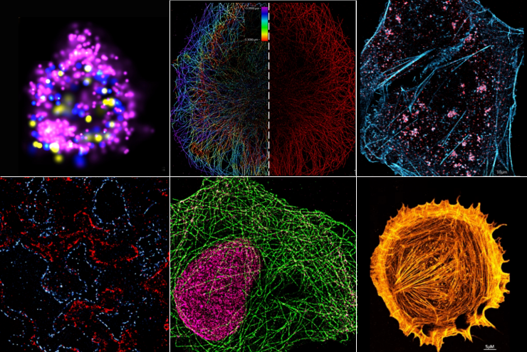
Super resolution microscopy
Techniques and illumination modes
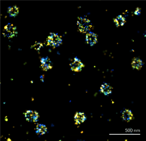
dSTORM & PALM microscopy
Achieve over 10-fold enhancement in resolution compared with widefield with dSTORM and PALM microscopy.
20 nm resolution
Resolve and visualize structures down to 20 nm in the XY plane.
3D imaging
Engage the astigmatic lens and explore the nanoworld in 3D with a Z resolution reaching 50 nm.
Quantitative results
Fluorescence microscopy becomes quantitative with dSTORM and PALM, count individual molecules as well as resolve structures.
Single-molecule FRET (smFRET)
Real-time nanoscale ruler operates on a 2-10 nm range. ONI is the only provider of commercial solutions for smFRET.
Insights into molecular interactions
Determine the spatial proximity of protein molecules so you can identify molecular interactions, binding events and dwell times.
Large field of view captures thousands of smFRET events
Increase experimental throughput by capturing thousands of single molecules at the same time.
Dedicated smFRET analysis
Plot and group individual FRET traces, as well as population averages.
smFRET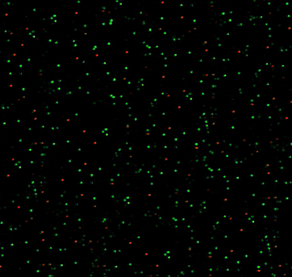
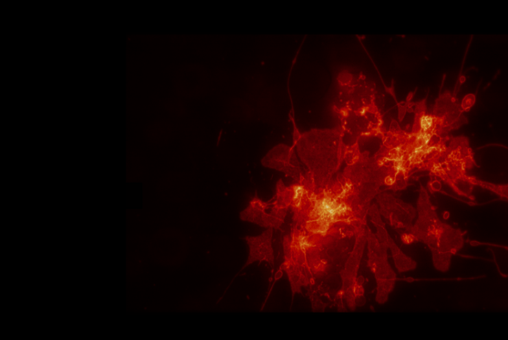
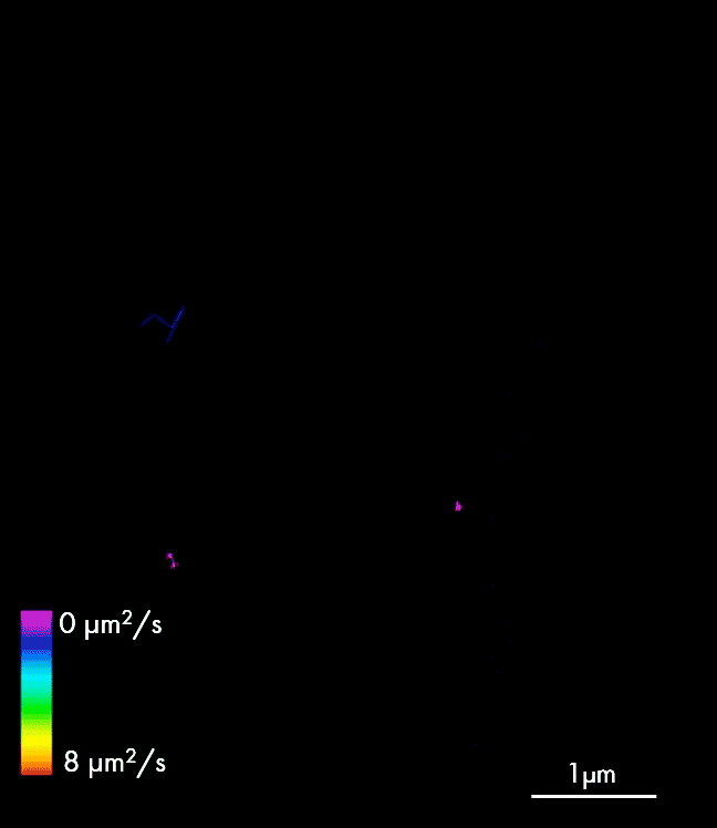
Single-particle tracking
Follow particles at a nanometer scale simultaneously in two channels.
Live cell imaging
With built in heating control, autofocus and support for microfluidic setups, live-cell imaging couldn’t be easier.
Track in four colors. Two at once
With four laser colors, you can track up to four different molecules in one experiment, with two tracked simultaneously.
Dedicated tracking analysis
Our dedicated NimOS software enables automatic particle tracking as well as measurement of diffusion coefficients, particle sizing and concentrations.
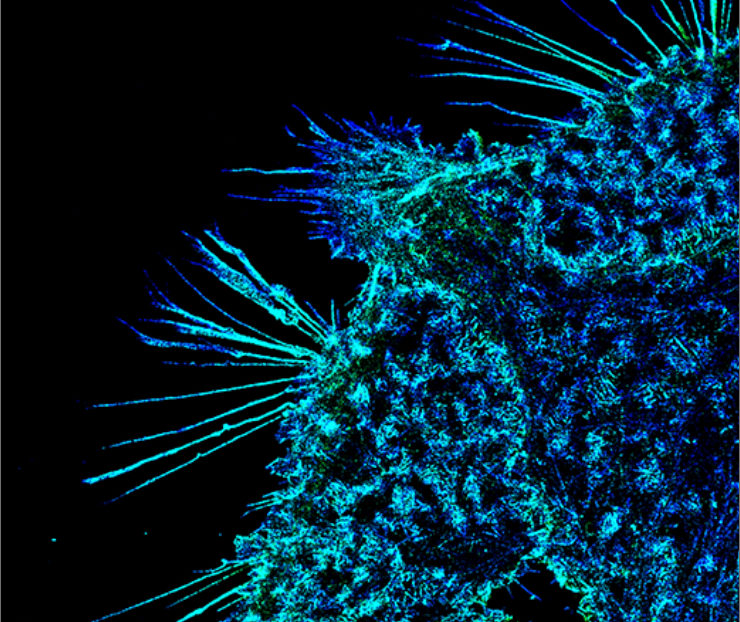
PAINT imaging
Transient binding for super-resolution imaging
20 nm resolution
Resolve and visualize structures down to 20 nm in the XY plane.
Higher localization precision
A longer blink means a brighter spot that can be detected by microscopes like the Nanoimager with higher localization precision, leading to images of higher resolution.
Reduction of signal blinking
In PAINT, fluorophores are constantly replaced by new imaging strands through binding kinetics, which prevents them from being permanently bleached like in dSTORM imaging where these are fixed to the epitopes.