Upcoming Chat With a Scientist – 30-minute one-on-one virtual discussions with ONI scientists book a session>
Super-resolution microscopy simplified
Where are you in your super-resolution journey?
ONI is here to support you along your super-resolution journey
Answer a few simple questions to discover key resources that can help you make the most of single-molecule imaging technology across a wide range of applications
I’m new to super-resolution
Welcome to the world of super-res!
How can you use super-resolution for your application or biological question?
Want to learn more about its uses?
I’m planning my experiment
Find what technique might be more suitable for your biological question
Key considerations for sample preparation
Want more technical and step-by-step details?
Choose your technique
What biological information do you need?
Choose your technique
What type of sample do you have?
Choose your technique
What’s the size of your structure of interest?
Choose your technique
Do you need multicolor imaging?
Your technique
Within the Nanoimager, TIRF imaging might provide you with the necessary information within a diffraction-limited context. SIM or Airyscan might also be sufficient in this case, you can get resolution down to 250 nm (SIM) and 350 nm (Airyscan).
For smaller structures, single-molecule localization techniques (SMLM) like dSTORM or PAINT would be highly recommended to obtain high precision results. If advanced microscopy is not available, expansion Microscopy could be used in this case.
For smaller structures, PALM imaging could be used here. PAINT is recommended in live cell mostly to quantify outer membrane structures via protein-protein interactions or weak interactions (uPAINT, glyco-PAINT, peptide-PAINT)
dSTORM imaging is the most suitable option in this case. It can achieve resolutions under 20 nm, allowing the visualization of structures and localization of molecules with great detail. It is compatible with multicolor imaging (up to 3) if needed.
PALM imaging is the most suitable option in this case. It can achieve resolution under 20 nm in both live or fixed samples.
DNA-PAINT might be the most suitable option in this case. It can achieve resolutions under 20 nm in both fixed or live sample, and is compatible with multicolor imaging. The lack of photobleaching due to continuously replenishing of fluorophores is also a benefit from PAINT.
Single Particle Tracking (SPT), usually combined with PALM, is the best option to follow the live dynamics of single molecules. Two-color SPT is also an available option.
STED for live cell imaging or in vivo STED for in vivo imaging can be helpful here. STED can reach temporal resolutions of < 50 ms
SIM for live cell imaging or in vivo SIM for in vivo imaging can be helpful here. SIm can reach temporal resolution of < 100 ns
smFRET seems the best option in this case, it enables distances between single molecules to be measured at a scale of 1-10 nm. Intra- and inter-molecular distances and stoichiometries can measured
PALM imaging could be an alternative to follow live molecular interactions, below 100 nm in axial resolution. For quantitative results, smFRET would be more suitable.
PALM imaging combined with Single Particle Tracking (SPT) could be an alternative to follow live molecular interactions, below 100 nm in axial resolution and for single molecules. For quantitative results, smFRET would be more suitable.
Unfortunately, we cannot find the technology you need. Please contact us for details
I’m ready to image
Double check you have everything you need
Master imaging techniques
Find your perfect microscope
I want to analyze my data
What are you looking to do?
Most single-molecule localization microscopy techniques, such as dSTORM, PAINT or PALM, allow the acquisition of high-resolution images (~20 nm) that provide morphological information of structures and the organization of proteins within them. For instance, to assess the distribution of a specific protein within a structure, a simple density profile measurement can be performed. 3D imaging and visualization further enhances the nanoscale information of structure morphology and protein organization.
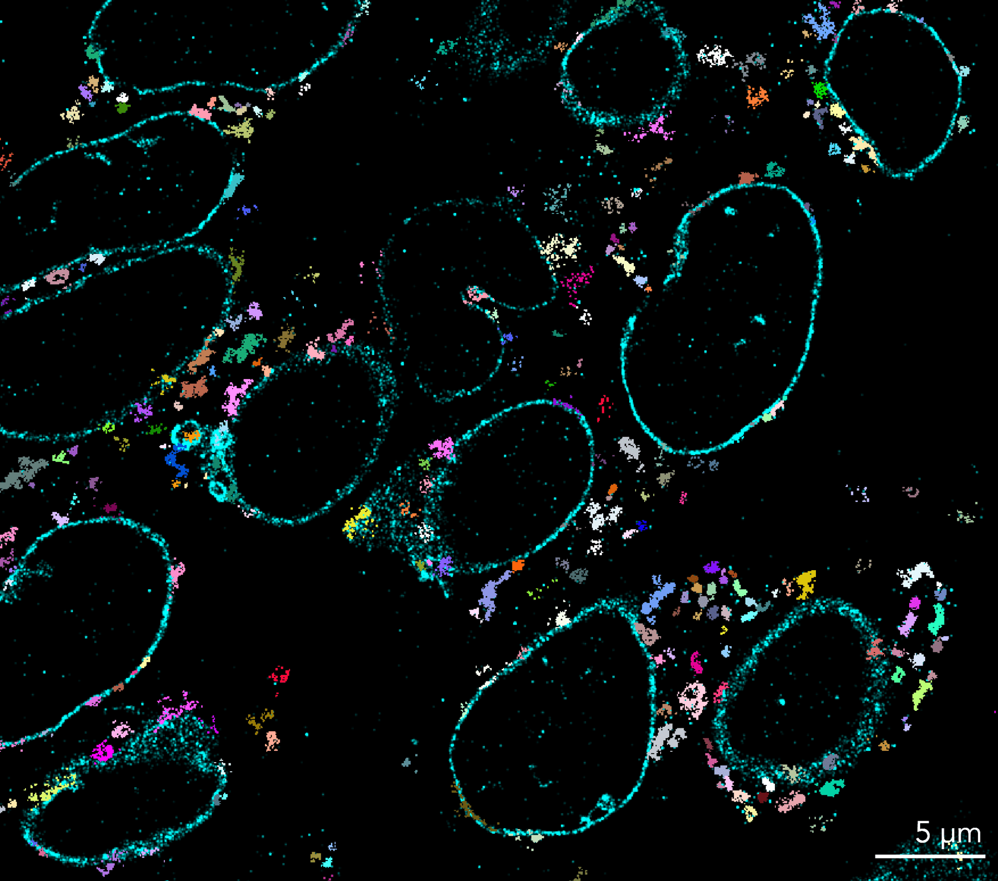
One of the most powerful applications of single molecule localization microscopy (SMLM) is investigating the spatial distribution of features down to the nanometer scale. The raw data obtained from SMLM experiments, such as dSTORM imaging, is a point cloud of localizations. This serves as the basis of nanoscale morphological analysis. We can use different clustering algorithms to detect the features of interest in the data. On ONI’s cloud-based CODI software, we offer different clustering analyses including DBSCAN, HDBSCAN and our in-house extension of HDBSCAN called Constrained clustering. Clustering on localization data allows researchers to find biological structures and quantify their morphological properties (size and shape), as well as determine presence/absence, even down to the level of expression, for specific markers of interest. You can find out more about clustering on this blog post.
The nanoscale resolution and clustering-based identification of structures allows investigation on the spatial relationship between structures and proteins. This powerful information can be extracted with different algorithms, such as K-Nearest Neighbors (KNN), which uses proximity to classify clusters. Please reach out to our team to find out more about using this and similar tools on our platform.
A critical part of SMLM imaging is to remove false positive localizations or noise to obtain accurate information and sensitivity for the analysis. To this end, the CODI analysis workflow includes a single-molecule filtering step that enables real-time visualization adaptation as users tune the filtering parameters.
You can use the SPT tracking functions within NimOS to detect fluorescent signals that form tracks. This allows users to follow biomolecule movement and extract particle speed and size distributions. For advanced analysis, Trackmate is a popular tool used by researchers.
PALM imaging coupled with Single Particle Tracking informs us how two molecules interact and behave over time in a live-cell environment. The NimOS tracking analysis allows a quick and in-depth analysis for the dynamic behavior of the biomolecules.
There are different ways to understand interactions between biomolecules. smFRET enables distance-based kinetic measurements between single molecules within a distance of 1-10 nm each, as well as intra- and inter-molecular distances and stoichiometries. The Nanoimager software NimOS enables smFRET analysis as soon as the acquisition has finished, allowing a quick evaluation to make a quick and informative decision.
The EV Profiling App on CODI is the perfect analysis workflow for EV samples with various subpopulations. The App can quantify size and biomarker distributions from a collection of single EVs. The App includes a range of tools from drift correction, single-molecule filtering, clustering, cluster filtering, and counting of biomarker distribution. In addition, the batch analysis tool allows analysis automation by applying the same analysis settings to multiple datasets.
Our cloud-based software platform called CODI stands for Collaborative Discovery. We are always happy to hear about features or tools that you would like to use for your applications.
Our CODI platform allows you to download images directly for publications with changeable color schemes, localisation sizes and contrast along with a scale bar. High-resolution image export is also available from NimOS, with file formats compatible with several third party softwares, such as FIJI ImageJ, Adobe Illustrator, and Affinity Designer.
Our CODI platform provides researchers with the possibility of sharing datasets, with different degrees of privacy (from close collaborators to fully public). This enables others to not only visualize datasets but also how they have been processed to reach the scientific conclusions. You can also explore our publications section on the website, with over 200 papers published with ONI products.
Our CODI platform provides researchers with the possibility of sharing datasets and analyses, and setting different levels of privacy as desired; thereby providing a one-end collaborative platform for the super-resolution community.
Unfortunately, we cannot find the technology you need. Please contact us for details
Products

Aplo Scope
A new era in super-resolution microscopy. Combine high-power SMLM imaging with low power live cell imaging across a expansive fully homogeneous FOV.
Now powered by CODI, Aplo Scope increases experimental throughput with 5 color imaging capabilities and offers maximal spectral discrimination.

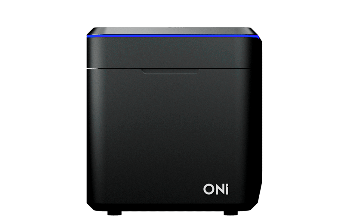
Nanoimager
Several super-resolution imaging modalities in one compact benchtop microscope. dSTORM, PALM, PAINT, Single-Particle Tracking, smFRET, TIRF, HILO… explore its possibilities for your research!

Aplo Flow
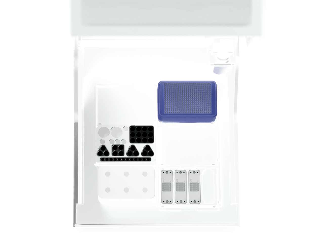
Application-specific and customizable sample preparation with fully automated liquid handler and the most user-centric, reliable, and reproducible end-to-end SMLM solution.

Kits
Start your complete super-resolution experience using our sample preparation kits to help you fix and label your samples: from cells to nanoparticles.

CODI
CODI is your cloud-based analysis platform, where you can collaborate with colleagues by sharing data and analysis workflows from anywhere.
ONI products
on CODI
& growing weekly
Gallery


FAQs
With unparalleled ease-of-use and simplicity, Aplo Scope has been designed to eliminate barriers to entry into the traditionally complex world of super-resolution microscopy and single-molecule imaging, and allow users of any skill level to utilize the instrument within one day of training.
The footprint of the microscope body is as small as a standard laptop 220 mm (w) x 421 mm (d) x 242 mm (h), the light engine measures 215 mm (w) x 420 mm (d) x 456 mm (h) and looks like a computer tower. They’re attached to each other by a 1.5 m long cable and, importantly, can be used on any bench or desk without needing a dark room or optical table.
Yes, samples can be stained with any super-resolution microscopy-compatible dye and imaged using Aplo Scope. However, this would require manual acquisition and analysis within CODI, or a third party application. ONI offers end-to-end kits where sample preparation, image acquisition, analysis and reporting are all integrated into a workflow, making imaging a seamless and more automated experience.