Enter now for your chance to win a 3 month Nanoimager subscription to study your EVs Enter here
Single Particle Tracking
A Simple Overview
What is single-particle tracking?
Single-particle tracking (SPT) is a powerful tool in microscopy that allows the motion of individual fluorescently-labeled particles (molecules, vesicles, virions or other molecular complexes) to be followed within a medium or in living cells and to obtain information on their dynamic behavior over time.
Single-particle tracks can help scientists better understand the positions, paths and interactions of molecules in systems that are highly dynamic or require imaging over extended periods of time. After data acquisition, the positions of the tracked particles are determined with nanometer-precision using image processing algorithms. Trajectories can also be extracted from time sequences with a resolution of up to milliseconds.
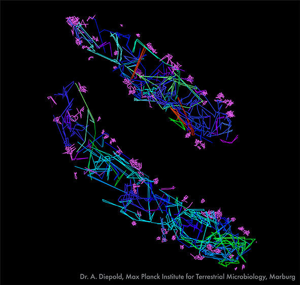
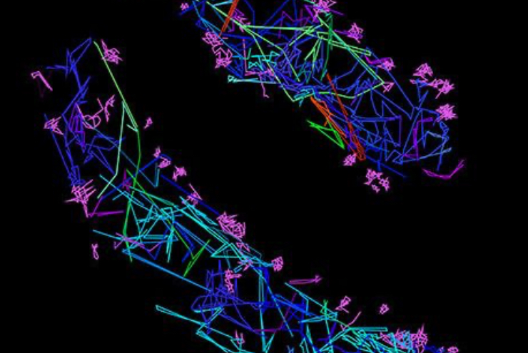
Single-particle tracking of T3SS-HALO stained with JF549 in live bacteria. Sample provided by Dr. A. Diepold, Max Planck Institute for Terrestrial Microbiology, Marburg, Germany. Scale bar is 1µm.
Why is single-particle tracking useful?
Single-particle tracking has been used to investigate a wide range of biological processes, including the motion of molecules (proteins or DNA) in the cytoplasm and nucleus, motion of vesicles or viral particles in the cytoplasm or medium, movement of lipids and proteins within membranes or across cellular compartments, and the uptake of external factors during gene or drug delivery, for instance. Single-particle tracks can also reveal structure-function relationships underlying complex systems and help generate 2D or 3D localization maps of such systems.
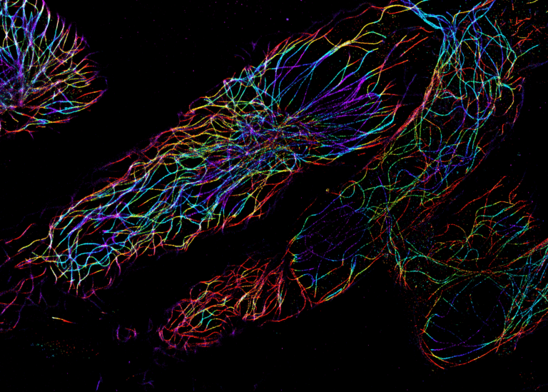
Atlastin1 protein walking along the ER in fibroblast cells, Sample provided by Dr. Christopher Obara, postdoc in Jennifer Lippincott-Schwartz lab, Janelia Farm
Why should I consider the Nanoimager for single-particle tracking?
Single-particle tracking in microscopy requires high speed and high sensitivity to achieve a time-resolution of milliseconds. The Nanoimager offers a unique single-molecule sensitivity that makes the detection of fluorescently-labeled molecules or vesicles trivial. With the Stage Top Incubator, live-cell imaging can be extended by maintaining optimal conditions for your samples. Our microscope uses single-particle tracking to both count and measure the size of molecules or vesicles in solution.
The Nanoimager can be heated to 37℃ for live-cell imaging and has four different laser lines. This offers the possibility of imaging two fluorophores simultaneously on a single sample and allows tracks registered in one channel to be assigned to cellular markers in the second channel. Our NimOS software provides a dedicated tracking analysis and automated detection of particles over time. The diffusion coefficients and measurements of the size and number of particles (for tracked particles in solution) is summarized instantly, for single or groups of tracks, with the option to export results in a convenient CSV format.
The Nanoimager has been used to image vesicles containing TRP channel proteins being transported to the plasma membrane, to track vesicles in cells and in solution in 3D, and to study the uptake of exosomes in HeLa cells.
Learn more about the different applications of the Nanoimager and the various super-resolution techniques that it supports.
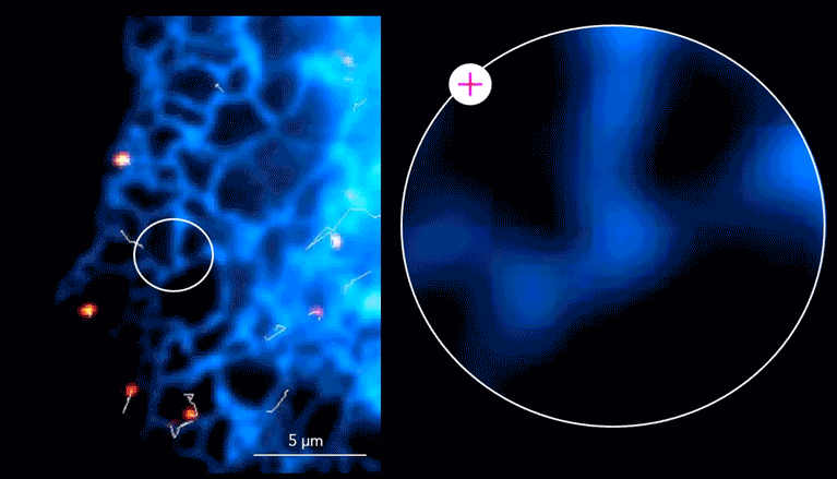
Zakrzewska-Czerwinska lab, Wroclaw University, Poland (scale bar 0.5µm color scale 0-6 um2/s)