<p><strong>Introducing Aplo Scope:</strong> A new era in super-resolution imaging! <strong><span style="text-decoration: underline;">learn more ></span></strong></p>

Introducing Aplo Scope: A new era in super-resolution imaging! learn more >

dSTORM

What is dSTORM Microscopy & why is it useful

dSTORM About How it works Usefulness Compatibility
About

Since its invention in 2006, STORM microscopy has become the most widely used super-resolution microscopy technique for single-molecule imaging. STORM stands for “Stochastic Optical Reconstruction Microscopy” and it relies on the stochastic activation of individual fluorophores with photoactivatable properties. STORM is compatible with fixed cells and fixed particles and requires specific antibodies. It can generate images of molecular structures resolved to under 20 nm.

How it works

During STORM, single fluorophores “blink” by a process of random activation from an off or dark state, to an on or emission state, quickly followed by a switch back to a dark state or photobleaching. This process is sequentially repeated many times until most fluorophores have been imaged. For a successful STORM imaging, individual fluorophores must be sparse enough, so that only one molecule is activated within a diffraction-limited region at any given time.

In the STORM reconstruction process, individual molecules can be precisely localized by determining their position coordinates from the photons detected for each activation event (often as a Gaussian function).

Usefulness

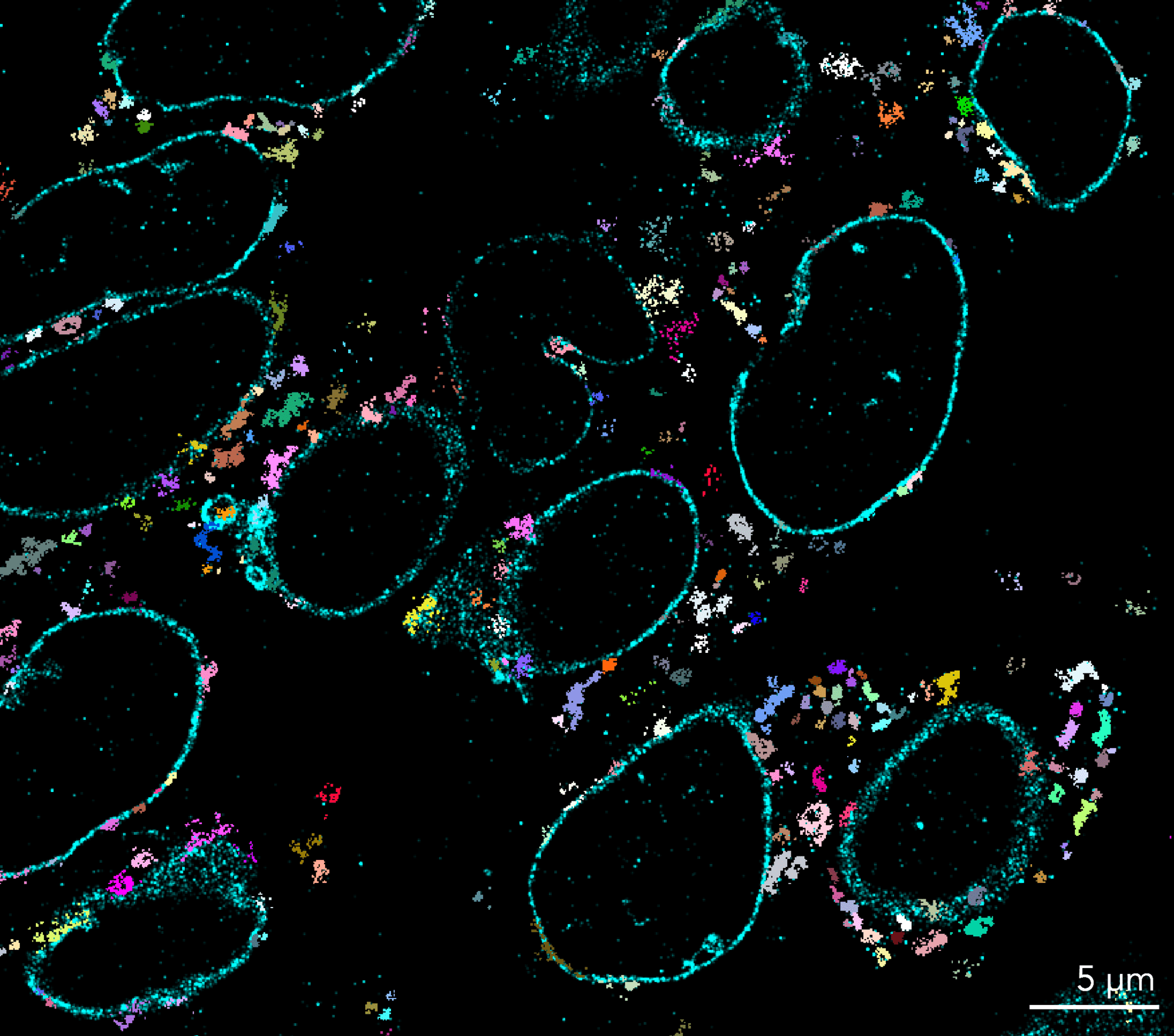
By stochastically imaging small subsets of photoswitchable fluorophores over time, STORM microscopy allows to spatially resolve the localization of individual molecules with high precision even in dense populations. STORM microscopes can produce high-quality images of cellular components resolved to under 20 nm, changing the way scientists visualize molecular structures, interactions within organelles and biological processes.

While STORM is an excellent localization-based super-resolution technique, its use for live imaging is limited, mainly due to phototoxicity issues. In this case, PALM microscopy, which uses different photoactivatable fluorophores, is better suited for live imaging and single-particle tracking. Equally, STORM requires specific antibodies for the target of choice, if such antibodies are not available, PALM can be an alternative.

Compatibility

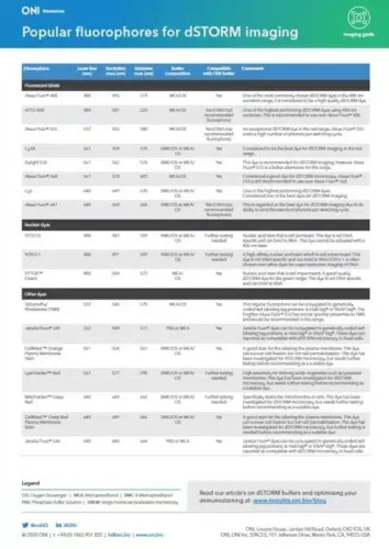
There are two variants of STORM. The first one, known as direct STORM or dSTORM microscopy, is compatible with many commonly used fluorophores, which can be converted to an off state using specific excitation parameters and in combination with specialized oxygen-scavenging imaging buffers. Ideal fluorophores for STORM should be very bright, have a high rate of photoswitching and exhibit minimal photobleaching in thiol-containing buffers. Some of the most common dSTORM fluorophores are Alexa Fluor™ and CFⓇ Dyes.

The second type of STORM microscopy uses a pair of activator-reporter dyes; the “activator” induces a switching and the “reporter” emits the signal detected. While the multiple activator-report pairs available allow multicolor STORM applications, this system requires dual labelling of the same target antibody and relies on the proximity of the two fluorophores. STORM without the activator dye, dSTORM, is currently widely used with different report dyes, as the photoswitching induced is efficient enough with substantial laser power.

How ONI can help in your dSTORM journey

Sample Preparation
Automation
Acquisition
Analysis
Sample Preparation
Automation
Acquisition
Analysis

Training Kit: dStorm

Designed to provide a simple workflow for new and existing users to learn the fundamentals of single-molecule localization microscopy

dStorm Discovery Kit

Provides a modular workflow for immunofluorescent labeling in cells with optimized dSTORM reagents

Aplo Flow

Application-specific and customizable sample preparation with fully automated fluidic control and the most user-centric, reliable, and reproducible end-to-end SMLM fluidics solution on the market today.

The Nanoimager

Several super-resolution imaging modalities in one compact benchtop microscope. dSTORM, PALM, PAINT, Single-Particle Tracking, smFRET, TIRF, HILO… explore its possibilities for your research!

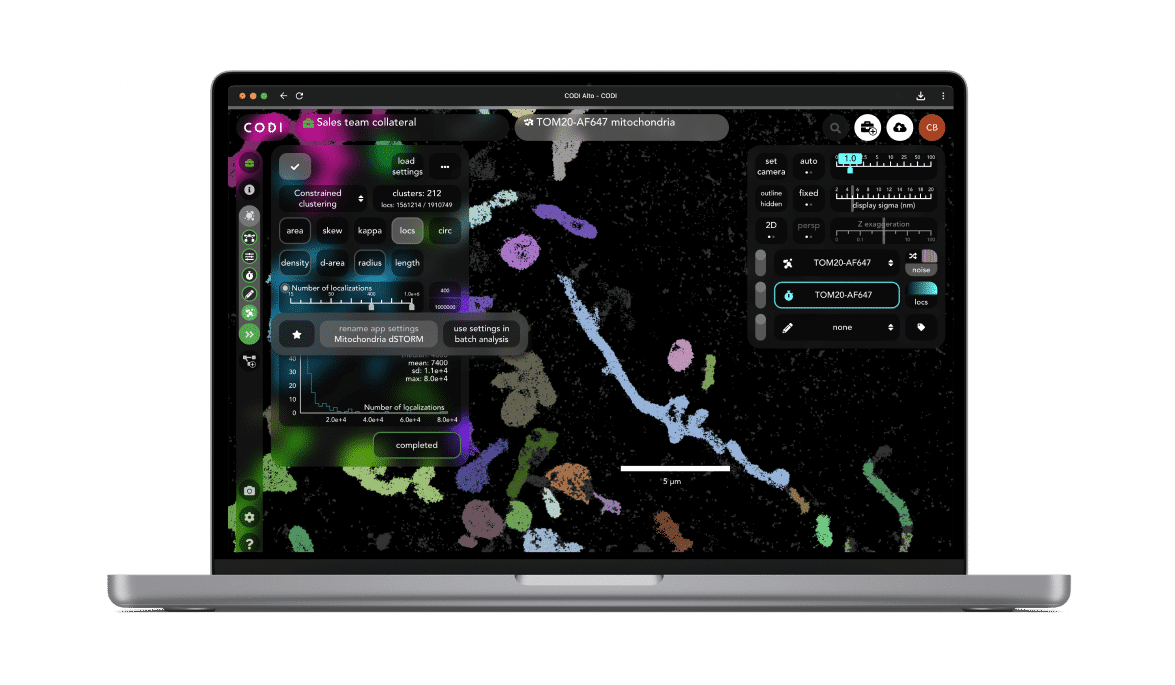
CODI

CODI is your cloud based analysis platform, where you can collaborate with colleagues by sharing data and analysis workflows from anywhere.

Important considerations

How do I choose my
fluorophore?
How do I choose my
fluorophore?
How do I choose my
fluorophore?
Publications
Technical Content
Videos
BiorXiv, January 2024
A nascent riboswitch helix orchestrates robust transcriptional regulation through signal integration,
Adrien Chauvier, Shiba Dandpat, Rosa Romero et al.
SCIENCE ROBOTICS, April 2022
A DNA molecular printer capable of programmable positioning and patterning in two dimension,
Erik Benson, Rafael Carrascosa Marzo, Jonathan Bath et al.
Journal of Extracellular Vesicles, April 2022
A functional corona around extracellular vesicles enhances angiogenesis, skin regeneration and immunomodulation,
Martin Wolf, Rodolphe W. Poupardin, Patricia Ebner-Peking et al.
Scientific Reports, March 2022
A coordinate-based co-localization index to quantify and visualize spatial associations in single-molecule localization microscopy,
Jelmer Willems , Harold D. MacGillavry
Methods, Volume 153, August 2018
A guide to nucleic acid detection by single-molecule kinetic fingerprinting,
Alexander Johnson-Buck, Jieming Li, Muneesh Tewari et al.
The Journal of Physical Chemistry B, December 2018
A Bayesian Nonparametric Approach to Single Molecule Förster Resonance Energy Transfer,
Ioannis Sgouralis , Shreya Madaan, Franky Djutanta et al.

Gallery

2 color dSTORM nucleus & mitochondria
CD63-tubulin dSTORM
Colorectal cancer tissue clusters
Tubulin dSTORM
Tubulin 3D dSTORM
Clathrin DNA-PAINT
2 color dSTORM nucleus & mitochondria
CD63-tubulin dSTORM
Colorectal cancer tissue clusters
Tubulin dSTORM
Tubulin 3D dSTORM
Clathrin DNA-PAINT