Enter now for your chance to win a 3 month Nanoimager subscription to study your EVs Enter here
Live-cell imaging
Simple Overview
What is live-cell imaging?
Live-cell imaging is a microscopy technique that allows in vivo imaging of cells, instantly and over a period of time. There are different types of microscopy compatible with live-cell imaging, which include both conventional contrast techniques, like differential interference contrast (DIC) or phase contrast, and fluorescence-based techniques.
The most commonly used are those capable of performing live-cell fluorescence microscopy, which allow researchers to study and follow the localization of fluorescence proteins in cells and in different tissues. Amongst these, scientists can use epifluorescence or widefield fluorescence, laser scanning or spinning disc confocal, lattice light-sheet and various super-resolution microscopy techniques, such as STORM or PALM.
A microscope adapted for live-cell imaging will include whole body heating or a heating device connected to the stage to keep cells at physiological temperatures and, when necessary, a CO2 incubator to support imaging over extended periods of time.
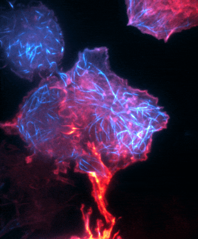
Murine macrophage RAW cells expressing Ensc-GFP (tubulin) and Ftractin-mCherry (actin). A TIRF timelapse was performed every 2 seconds. Sample provided by Dr. Valentim Jamouille from Clare Waterman’s lab at the NIH National Heart, Blood & Lung Institute, Bethesda (USA). Scale bar is 5µm.
Why is live-cell microscopy useful?
Live-cell imaging can provide a large amount of information and detail about a molecule of interest, cellular organelle or cell type within a tissue. Imaging of fixed samples, like those labeled with immunofluorescence staining, often only provide a snapshot of a specific cellular process or the localization of a molecule. This can limit the interpretation of information, particularly when studying highly dynamic processes.
With live-cell microscopy techniques, scientists can obtain highly valuable information of such biological processes, including:
• Molecular localization and movement with time resolution
• Dynamic complex assembly and interaction between molecules
• Organization of components within cells
• Vesicle or viral particle behavior
• Cellular responses to environmental cues
Can I do live-cell imaging on the Nanoimager?
The Nanoimager allows whole body heating and adjustable temperature settings, including 37°C and up to 42°C, to support live-cell imaging of different cell types in various organisms. Its compact design, unrivaled stability and smart alignment design, makes it a robust microscope that can be used in any laboratory environment.
The Nanoimager offers powerful imaging of single molecules with enhanced resolution of up to 20 nm by supporting different super-resolution techniques, including dSTORM, PALM, single-particle tracking and smFRET. smFRET provides quantitative information on dynamic protein-protein interactions. PALM microscopy offers even better resolution and can be combined with HILO or TIRF microscopy to improve the signal to noise ratio of thinner samples.
The Nanoimager’s NimOS software includes a single-particle tracking feature, which provides tracking analysis, particle size and number measurements, and automated detection over time. Our microscope can image two fluorophores simultaneously (with four laser colors) on a single sample, allowing tracks registered in one channel to be assigned to cellular markers in the second channel.
Learn more about the Nanoimager’s super-resolution microscopy techniques and applications.
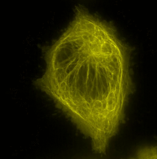
SIM imaging done on live cells: Hel Cells stained with GFP-Tubulin. Sample provided by Steve Thomas and Natalie Poulter; bar: 10 um

