Upcoming Webinar – Automated DNA-PAINT and Exchange Imaging with Aplo Flow Fluidics register now>
CODI
Acquisition, analysis and reporting in one place

Intuitive cloud-based solutions
Sample to answer
CODI is a comprehensive software solution with automated image acquisition, analysis and reports anyone can use
Storage and processing
CODI runs on the cloud and offers unlimited storage, so you can focus on data analysis and not computer requirements
Analysis & reporting simplified
Analysis app workflows can be customized to analyze hundreds of datasets and generate reports with ease
Sharing and collaboration
CODI datasets and analyses can be organized into collaborations and instantly shared with collaborators
CODI unifies the Aplo Platform

Aplo Scope
Effortless super-resolution and live-cell imaging
CODI powers Aplo Scope – the flagship innovation behind the Aplo Platform, a versatile easy-to-use microscope designed for reliability and maximal performance across experiments and users of any skill level.
Aplo Scope Control is built into CODI, allowing full control of the microscope’s advanced hardware while integrating with CODI’s powerful data organisation and analysis tools. Intuitively perform up to 5 color panels, fixed and live-cell imaging.

CODI
Analyze and share data instantly
Automatically upload and analyze your data using CODI’s powerful cloud analysis workflows, which simplify single molecule data analysis. Save custom analysis settings to specific experiments, then apply them to newly acquired datasets for experimental reproducibility.
CODI datasets and analyses can be organized into collaborations, shared and analyzed from any computer. Easily find, share, and export images and reports for publication.

Aplo Flow
Fluidic automation to increase throughput
Aplo Flow combines application-specific sample preparation with fully automated fluidic control, enhancing the speed and efficiency of super-resolution microscopy.
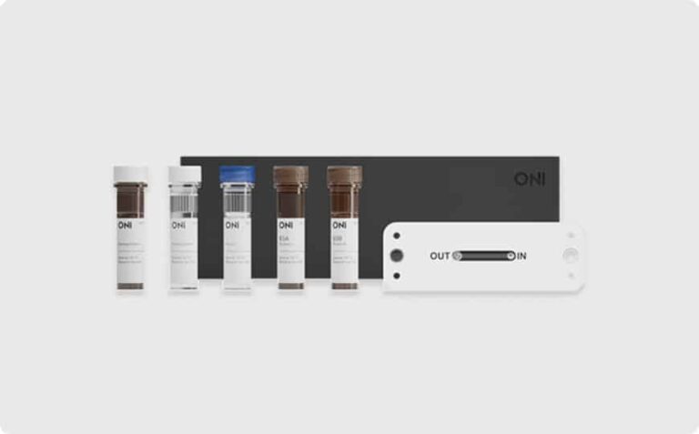
For Extracellular Vesicle sample prep with EV Profiler 2, CODI mimics the layout to the assay chip and the Aplo Flow deck, easily assigning reagents to lanes. Metadata including sample names and staining details are automatically transferred from acquisition through to AutoEV o
What’s in CODI


With CODI you are able to
Understand the morphology of my structure

Single-molecule localization microscopy techniques, such as dSTORM, PAINT or PALM, pinpoint the position of individual molecules, providing morphological information of structures and the organization of proteins. To assess the distribution of a specific protein within a structure, a simple density profile measurement can be performed. 3D imaging and visualization further enhances the nanoscale information of structure morphology and protein organization.
Understand how my molecules of interest organize

One of the most powerful applications of SMLM is investigating spatial distributions down to the nanometer scale. The raw point cloud of localizations serves as the basis of nanoscale morphological analysis. CODI offers different clustering analyses including DBSCAN and our in-house extension of HDBSCAN called Constrained Clustering. Clustering on localization data allows researchers to find biological structures and quantify their morphological properties (size and shape), as well as determine presence or absence of specific markers of interest.
Clean up my data to improve signal-to-noise ratio

A critical part of SMLM imaging is removing noise to obtain accurate information and sensitivity for the analysis. To this end, the CODI analysis workflow includes a single-molecule filtering step that enables real-time visualization as users tune the filtering parameters, as well as default filters that serve as a great starting point for keeping only the most accurate data.
Identify EV subpopulations based on size
or biomarker content

The EV Profiling App on CODI is the perfect analysis workflow for EV samples with various subpopulations, allowing you to quantify size and biomarker distributions from a collection of single EVs. The App includes a range of tools from drift correction, single-molecule filtering, clustering, cluster filtering, and counting of biomarker distribution. In addition, the batch analysis tool allows analysis automation by applying the same analysis settings to multiple datasets.
CODI Apps
CODI contains several app workflows to streamline and simplify single molecule data analysis. Each workflow is a unique combination of tools for a specific kind of dataset, all starting with the basics of spatial drift correction and single-molecule localization filtering.
Clustering

The Clustering App provides all of the necessary analysis widgets for cell-based clustering analysis workflow.
Key outputs:
- Real-time data visualization, including histograms of various cluster metadata
- Clustering CSV containing quantitative and morphological information about each cluster
- High-resolution images of single molecule localization and their assigned clusters
Clustering & Counting

The Clustering & Counting App provides a complete single-molecule localization microscopy analysis workflow. It includes all the widgets available on Clustering App and a Counting widget.
Key outputs:
- Real-time data visualization, including histograms of various cluster metadata
- Clustering CSV containing quantitative and morphological information about each cluster
- High-resolution images of single-molecule localization and their assigned clusters
- Summary report segmenting clusters into user-defined phenotypes
EV Profiling

The EV Profiling App is the perfect analysis workflow for Extracellular Vesicles (EV) datasets. It contains a set of algorithms customized for quantifying size and biomarker distributions of single EVs in a sample.
Key outputs:
- Real-time data visualization, including histograms of various EV metadata
- Clustering CSV containing quantitative and morphological information about each EV
- Summary report segmenting EVs into user-defined phenotypes
- High-resolution images of EVs and their single molecule localizations
CODI Support
FAQs
Do you have to pay to use CODI?
No. Everyone using a Nanoimager gets access to CODI and its applications at no charge during the Beta phase of release.
Can CODI analyze data acquired on other microscopes?
Technically it can, however, it is currently designed to support data acquired from the Nanoimager.
Do you require a high-specification computer to run CODI or the Nanoimager software?
No. CODI fully runs on the cloud, so you can do your analysis anywhere. The software to run the Nanoimager (currently NimOS) works on most modern computers, running 64-bit Windows 10. We supply a high-specification laptop with each Nanoimager and this is included in the price.