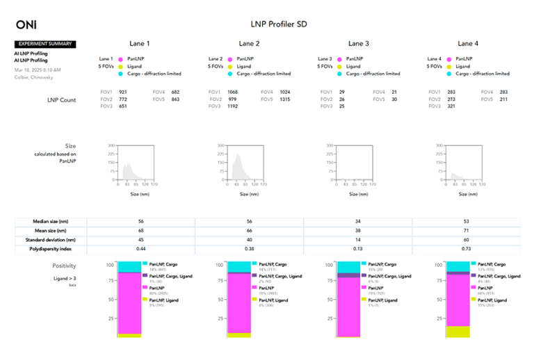
<p><strong>BrainSTORM Week</strong>: Register now for our upcoming dSTORM webinar series! <strong><span style="text-decoration: underline;">learn more</span></strong></p>

BrainSTORM Week: Register now for our upcoming dSTORM webinar series! learn more

  • Applications

Lipid nanoparticles

Lipid nanoparticles (LNPs) are advanced delivery systems formed from tiny, spherical lipid structures. They can encapsulate various therapeutic agents, including drugs and genetic materials like mRNA, protect them from degradation and facilitate their entry into target cells. By merging with cell membranes, these nanoparticles ensure effective delivery and release of their cargo, making them crucial for innovations in mRNA vaccines, gene therapies, and personalized medicine.

Overview

LNPs Significance Challenges Trends Tools
Significance

Significance

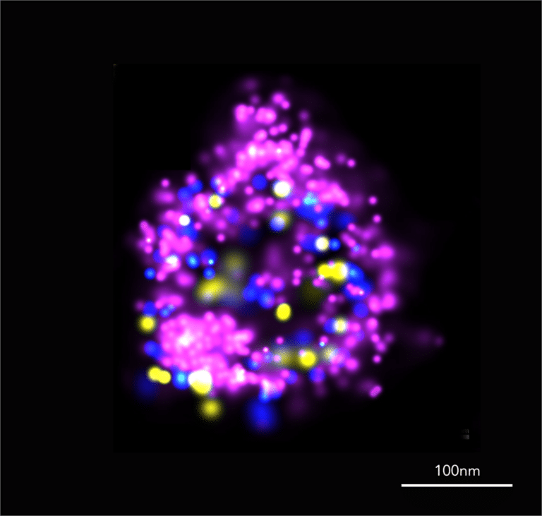
LNPs have transformative therapeutic significance due to their ability to deliver nucleic acids, such as mRNA and RNA-based drugs, with high efficiency and specificity. Since the approval of the first LNP-based therapy, Onpattro, in 2018 and their global explosion in 2020, they revolutionized drug discovery and development, and nowadays new therapies are developed at an unprecedented speed and efficiency.

The LNP lipids encapsulate sensitive therapeutic agents and protect them, facilitating their delivery into target cells with minimal degradation.

Challenges

Challenges

Some of the challenges in LNP-based therapeutics are due to the low yield of LNPs with the required size, cargo loading and ligand density, which are essential for their performance. Without information on these features, determining the stability, efficiency and specificity of the LNPs is extremely difficult. Obtaining reliable, reproducible data at a single LNP level paves a path to optimized production of high-quality therapeutic agents.

Trends

Trends

Recent trends in LNP research focus on enhancing delivery precision and expanding therapeutic applications. Advances in lipid chemistry are leading to the development of more sophisticated LNP formulations with improved stability, lower toxicity, and better targeting capabilities. Researchers are also exploring personalized medicine approaches, using LNPs to deliver tailored treatments for specific genetic profiles or disease conditions.

Additionally, there is growing interest in combining LNPs with other technologies, such as CRISPR and RNA-interference, to address complex diseases more effectively. These trends reflect a broader push toward maximizing the versatility and efficacy of LNP-based therapies.

Tools

Tools

Current tools used for LNP research include a range of advanced techniques that enable precise characterization and optimization of these delivery systems. High-performance liquid chromatography (HPLC) and dynamic light scattering (DLS) are commonly employed to analyze LNP size, distribution, and composition. Mass spectrometry can also be used to monitor LNP stability and content.

Cryo-electron microscopy (cryo-EM) provides detailed structural insights at the nanoscale, while super-resolution microscopy offers a quantifiable and detailed image within the cellular environment: single-molecule localization microscopy (SMLM) offers enhanced visualization and in-depth characterization of LNPs in physiological-like conditions, and nanoparticle tracking analysis (NTA) is used to assess sizing and stability, both surpassing the limits of conventional optical microscopy.

Why super-resolution for LNP research?

The ONI Nanoimager enables users with a complete set of tools to characterize LNPs and understand their cellular uptake in a single platform capable of generating unique insights to never before seen through other analysis methods.

Super-resolution insights into LNPs

Single LNP analysis
LNP structure
Cargo loading
Ligand loading
Single LNP analysis
LNP structure
Cargo loading
Ligand loading

Single LNP analysis

With super-resolution microscopy, analyzing individual LNPs has never been easier. This technique offers high-detail imaging in physiological-like conditions, enabling clear visualization and distinction of individual LNPs, even when they are close together.

Our Nanoimager offers simultaneous analysis of thousands of individual LNPs, at a single LNP level. This enhanced clarity provides critical insights into the loading efficiency of both cargo and ligand, and allows scientists to optimize their production processes to maximize and perfect their yield.

Figure (left): SMLM offers quantitative insights into LNP characteristics, including accurate sizing, detection of cargo and efficiency of ligand loading, at a single LNP level.

Morphology, Size and Stability

Single Molecule Localization Microscopy (SMLM), specifically dSTORM, provides a powerful tool for examining the morphology of individual lipid nanoparticles (LNPs) with unprecedented resolution.

Unlike techniques like flow cytometry and mass-spectrometry, which aggregate data, dSTORM provides precise nanometer-level details on LNP morphology and size. It is crucial for quality control to measure size variability accurately: small particles may indicate degradation, large ones could be aggregates, and high polydispersity suggests instability. Thus, assessing size variability helps evaluate sample quality.

With the full information on size variations and structural anomalies, scientists can accurately assess their LNP behavior, and optimize the formulations for effective therapeutic applications.

Figure (left): Size distribution of two distinct LNP populations, measured with SMLM. The LNP Surface marker is in magenta.

Cargo loading

One of the key challenges in LNP development for therapeutics is to have the cargo encapsulated in as many LNPs as possible. For that, it is essential to assess how many LNPs contain cargo and under what conditions.

By combining SMLM imaging of the LNP surface marker, fluorescence imaging of the cargo, and advanced software tools, scientists can quantify their cargo positivity at a single LNP level. This valuable information attests to the loading efficiency without compromising on inaccuracies like signal averaging.

Figure (left): Size distribution of 3 LNP populations: Top row – empty LNP, middle row – cargo-containing LNP, bottom row – 1:1 mix of empty LNPs and cargo-containing LNPs.
Surface marker is in magenta, RNA cargo is in cyan (diffraction limited images).

Ligand loading

Drug delivery using LNPs is most effective when the LNP surface is loaded with ligands that direct the LNPs to specific target cells. Super-resolution microscopy encompasses the ability to inspect individual LNPs and measure ligand positivity and abundance accurately and reproducibly.

The abundance of ligands on each individual LNP correlates with targeting efficiency, so quantifying the ligand occurrence using SMLM is very valuable in assessing the effectiveness of ligand loading, alongside the ligand interactions with its targets. Gaining data on both the fraction of ligand positivity and its abundance provides the researcher with information on individual LNPs, as well as on their entire population.

Figure (left): Montage of representative SMLM LNP images. Surface marker is in magenta, ligand is in yellow.

Upgrade your LNP research with ONI

Nanoimager
CODI
Nanoimager
CODI

Nanoimager

Discover the potential of your LNP research with the Nanoimager, our benchtop microscope offering advanced super-resolution techniques like dSTORM, PALM, and Single-Particle Tracking. Experience unparalleled clarity and detail, allowing you to explore LNP size, morphology, and interactions with exceptional precision.

CODI

CODI is our cloud-based analysis platform, where you can collaborate with colleagues by sharing data and analysis workflows from anywhere.

Featured publications

3D-Printed LNP Production
IL-4 LNP Treatment
Drug Nanostructures in NLCs
LNP-EV Fusion

Rapid production of nanoscale liposomes using a 3D-printed reactor-in-a-centrifuge: Formulation, characterisation, and super-resolution imaging

This paper describes a new method to produce LNPs using a 3D-Printed Reactor-In-A-Centrifuge. The scientists used dSTORM imaging with the Nanoimager to demonstrate that their LNP have a consistent size and shape.

He, Y.; Grandi, D.D.; Chandradoss, S.; LuTheryn, G.; Cidonio, G.; Nunes Bastos, R.; Pereno, V.; Carugo, D. Rapid Production of Nanoscale Liposomes Using a 3D-Printed Reactor-In-A-Centrifuge: Formulation, Characterisation, and Super-Resolution Imaging. Micromachines 2023, 14, 1763. https://doi.org/10.3390/mi14091763

Resolving sepsis-induced immunoparalysis via trained immunity by targeting interleukin-4 to myeloid cells

Sepsis is a life-threatening condition that is characterized by both excessive inflammation, and, paradoxically, a subtype of immune suppression, called immunoparalysis. The cytokine IL-4 was shown to reverse the immunosuppressive phenotype in cultured cells, but it is not very effective when given as a drug on its own. The researchers engineered a version of IL-4 which can be integrated into LNPs that specifically target white blood cells of the innate immune system.

Schrijver DP, Röring RJ, Deckers J, de Dreu A, Toner YC, Prevot G, Priem B, Munitz J, Nugraha EG, van Elsas Y, Azzun A. Resolving sepsis-induced immunoparalysis via trained immunity by targeting interleukin-4 to myeloid cells. Nature Biomedical Engineering. 2023 Sep;7(9):1097-112. https://doi.org/10.1038/s41551-023-01050-0

Determination of nanostructures and drug distribution in lipid nanoparticles by single molecule microscopy

This paper uses super-resolution methods to model the spatial distribution of drugs within LNPs. They use both single-molecule tracking and single-molecule localization to define substructures within the LNP where their drug-like molecule is accumulating. Using these techniques they can demonstrate how encapsulation looks in 3D, therefore gaining important information on the efficiency of the production and possibly a prediction of the success of drug delivery. 

Boreham A, Volz P, Peters D, Keck CM, Alexiev U. Determination of nanostructures and drug distribution in lipid nanoparticles by single molecule microscopy. European Journal of Pharmaceutics and Biopharmaceutics. 2017 Jan 1;110:31-8. https://doi.org/10.1016/j.ejpb.2016.10.020

Programmable RNA Loading of Extracellular Vesicles with Toehold-Release Purification

LNPs are effective drug carriers, however, some of them can induce side-effects resulting from immune response against components of the LNPs. Extracellular vesicles (EVs) are naturally occurring lipid vesicles, therefore not triggering immune response. The researchers developed a method to fuse LNPs with EVs, in order to introduce the LNP cargo into EVs. They used the Nanoimager to monitor the fusion and to detect the cargo loading of the EVS.

Malle, M.G.; Song, P.; Löffler, P. M. G.; Kalisi, N.; Yan, Y.; Valero, J.; Vogel, S.; Kjems, J. Programmable RNA Loading of Extracellular Vesicles with Toehold-Release Purification. Journal of the American Chemical Society. 2024: 146 (18), 12410-12422. DOI: 10.1021/jacs.3c13123

Upcoming virtual events

Live virtual LNP demo

October 10, 2024

Discover the Nanoimager’s transformative impact on LNP research and see how it can enhance your studies—register for ONI’s Live Virtual Demo to experience its capabilities firsthand.

Webinar - Why SMLM for LNP research?

November 20, 2024

Enhance your LNP research with super-resolution microscopy. Join us for the webinar, “Why SMLM for LNP Research?” and explore how SMLM can provide critical insights and breakthroughs.

Back to top

Schedule your LNP demo

Gallery

AF488 labelled EV
CD63-tubulin dSTORM
Colorectal cancer tissue clusters
Tubulin dSTORM
Tubulin 3D dSTORM
Clathrin DNA-PAINT
AF488 labelled EV
CD63-tubulin dSTORM
Colorectal cancer tissue clusters
Tubulin dSTORM
Tubulin 3D dSTORM
Clathrin DNA-PAINT

FAQs

How can ONI’s Nanoimager enhance my LNP research?

ONI’s Nanoimager provides advanced super-resolution microscopy that allows for high-resolution imaging of lipid nanoparticles at the nanoscale. This capability enables detailed visualization of LNPs and their interactions within cells, improving the understanding of their behavior, distribution, and efficiency. By revealing finer details that traditional microscopy cannot, the Nanoimager supports more precise characterization and optimization of LNP formulations.

What advantages does ONI’s technology offer for analyzing LNP stability?

ONI’s technologies offer critical insights into LNP stability by allowing real-time observation of nanoparticles in various conditions. With high-resolution imaging and tracking capabilities, researchers can monitor changes in LNP structure and behavior over time, helping to identify factors affecting stability and making it easier to optimize formulations for better performance.

How can ONI’s tools assist in overcoming challenges in LNP targeting and delivery?

ONI’s tools provide enhanced visualization of LNP interactions with target cells and tissues. This detailed imaging helps in understanding how LNPs are distributed and taken up by specific cells, allowing researchers to refine targeting strategies and improve the efficacy of LNP-based therapies. By offering insights into cellular uptake and localization, ONI’s technology aids in optimizing delivery mechanisms for better therapeutic outcomes.

Subscribe to our mailing list