Upcoming Webinar – Small dyes, big impact: Enabling gentle super-resolution imaging in live cells register now>
Extracellular Vesicles
Exosomes and other extracellular vesicles (EVs) play key roles in cell-to-cell communication. EVs can cross biological barriers (such as the blood-brain barrier) and get internalized into the cell with a high degree of specificity. Thus, they are an ideal candidate for novel drug delivery methods and disease diagnostics.
About Extracellular Vesicles
Challenges in visualizing extracellular vesicles
A substantial and growing body of evidence highlights extracellular vesicles as critical components in cell-to-cell communication pathways. The visualization of EVs is fundamental to our understanding of the role of EVs in all aspects of cellular transmission; from the packaging of signaling molecules and nucleic acids during vesicle biogenesis, to tracking of their uptake and fate after internalization within selected target cells or tissues.
Characterize EVs with super-resolution
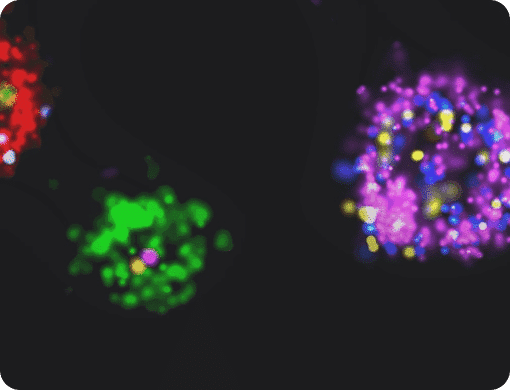
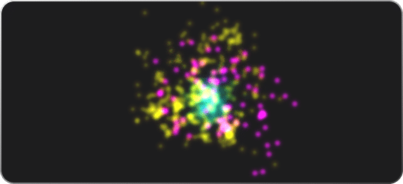
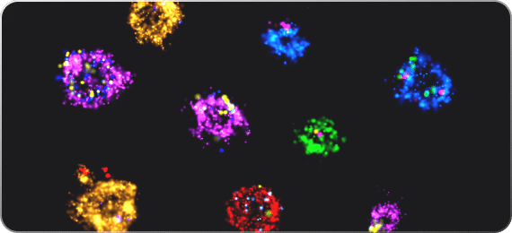
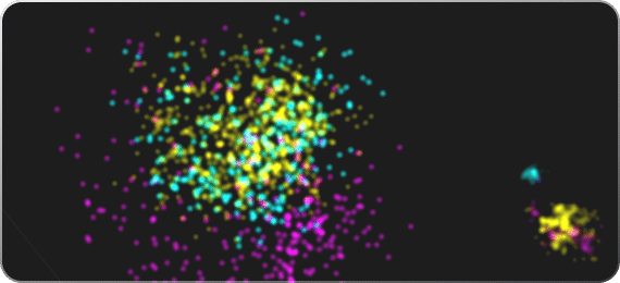
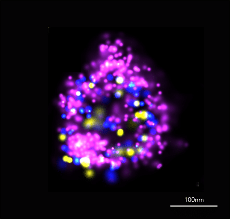
Super-resolution microscopy can be used to visualize and identify EVs in cells and in solution. Biomarkers can be stained in single EVs, each with a different fluorescence signature. By using one generic marker of EVs (like ONI’s Pan-EV stain) in one color and specific markers for EVs (such as CD9, CD63 and CD81) in others, it is possible to study the fraction of EVs with a particular biomarker within a population, confidently gaining insights on both single EVs and on the population.
Size, count, image… understand
One of the most challenging aspects of studying EVs, and perhaps one of the most important, is combining multiple complementary characterization techniques. Single-molecule fluorescence microscopy offers the most sensitive fluorescence measurements available, allowing us to extract information with greater sensitivity than with any other tracking-based fluorescence instrument.
Case Studies
ATP1A3 as a target for isolating neuron-specific extracellular vesicles from human brain and biofluids
In this paper, the scientists aimed to investigate the potential of the protein ATP1A3 to serve as a biomarker for Alzheimer’s Disease progression in EVs isolated from neurons and from liquid biopsies. They used a number of methods to identify ATP1A3 as an EV biomarker specific to neurons. Additional single EV analysis reveals that ATP1A3 is specific and abundant in induced neurons from humans, and its levels in EVs correlate with levels of Alzheimer’s Disease-related proteins like Aβ. Since ATP1A3 is abundant in EVs derived from liquid biopsies, it can serve as a marker for neurodegenerative diseases.
Yang You et al. ,ATP1A3 as a target for isolating neuron-specific extracellular vesicles from human brain and biofluids.Sci. Adv.9,eadi3647(2023). DOI:10.1126/sciadv.adi3647
Extracellular vesicles from the lung pro-thrombotic niche drive cancer-associated thrombosis and metastasis via integrin beta 2
In this work, scientists set out to explore the mechanism behind thrombosis in cancer patients. They discovered a subpopulation of the immune cell macrophages that reside in the lungs and secrete small EVs that are enriched with the protein Integrin beta 2. This protein binds glycoproteins on the surface of platelets and coagulation proteins in the blood, initiating thrombosis. This work included single EV analysis on clinical samples from patients, and is an important first step in identifying at-risk patients, as well as in developing treatments.
Lucotti, S. et al. Extracellular vesicles from the lung pro-thrombotic niche drive cancer-associated thrombosis and metastasis via integrin beta 2. Cell 188, 1642-1661 (2025), DOI: 10.1016/j.cell.2025.01.025.
Selenoprotein P is a target for regulating extracellular vesicle biogenesis and secretion from activated microglia in vivo
The researchers set out to study how the protein Selenoprotein P (sepp1) is involved in the regulation of EV secretion by microglia, the immune cells that guard the brain. They confirmed that sepp1 is important for normal EV production, and that its absence is affecting multiple EV characteristics, as well as other cellular pathways. Since EVs from microglial cells were shown to be involved in several neurological conditions, sepp1 can be a new drug target in patients.
Bodart-Santos V, Ruan Z, Melvin BC, Pandey I, Ikezu S, Ikezu T. Selenoprotein P is a target for regulating extracellular vesicle biogenesis and secretion from activated microglia in vivo. Cell reports. 2024 Dec 24;43(12), DOI: 10.1016/j.celrep.2024.115025
End-to-end EV workflow

Aplo Scope
A new era in super-resolution microscopy. Combine high-power SMLM imaging with low power live cell imaging across a expansive fully homogeneous FOV. Now powered by CODI, Aplo Scope increases experimental throughput with 5 color imaging capabilities and offers maximal spectral discrimination.

The Nanoimager
Discover the power of advanced super-resolution imaging with our compact benchtop microscope, designed to elevate your EV research. Featuring cutting-edge modalities like dSTORM, PALM, PAINT, Single-Particle Tracking, smFRET, TIRF, and HILO, this versatile system offers unparalleled imaging capabilities to explore and analyze extracellular vesicles in unprecedented detail.
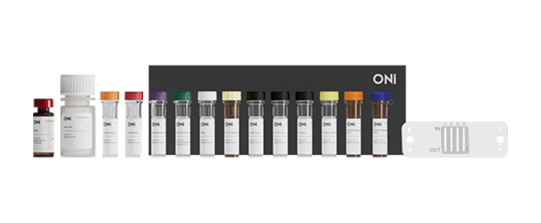
EV Profiler 2
ONI Application Kit™: EV Profiler 2 is ONI’s advanced reagent kit for visualizing and phenotyping extracellular vesicles with dSTORM microscopy on the Nanoimager, offering enhanced reproducibility, improved EV capture efficiency, and precise sizing and colocalization of up to three biomarkers per EV, complemented by AutoEV software for automated, comprehensive analysis in just 90 minutes.
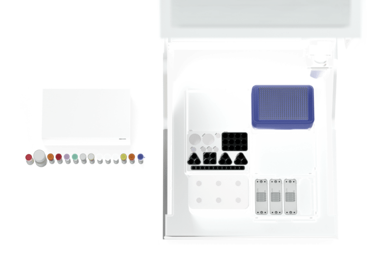
Aplo Flow
Achieve precision in EV research with our fully automated, customizable fluidics solution. Designed for reliable, reproducible sample preparation, it’s the most user-centric, end-to-end tool for super-resolution microscopy in extracellular vesicle studies.
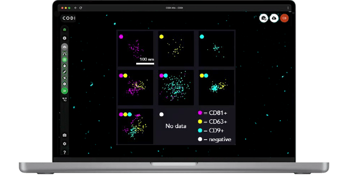
AutoEV in CODI
When combined with our newest software offering, AutoEV, your system will be calibrated and optimized to acquire and analyze a 4-lane chip automatically and deliver a comprehensive report of EV size and positivity for each individual lane in 90 mins.
Key Resources
Cell reports medicine, March 18, 2025
Single extracellular vesicle detection assay identifies membrane-associated α-synuclein as an early-stage biomarker in Parkinson’s disease,
Cell, February 11, 2025
Extracellular vesicles from the lung pro-thrombotic niche drive cancer-associated thrombosis and metastasis via integrin beta 2,
Cell Reports, December 24, 2024
Selenoprotein P is a target for regulating extracellular vesicle biogenesis and secretion from activated microglia in vivo,
Extracell Vesicles Circ Nucleic Acids,
Detection by super-resolution microscopy of viral proteins inside bloodborne extracellular vesicles.,
Journal of Extracellular Vesicles,
Exhaled breath condensate contains extracellular vesicles (EVs) that carry miRNA cargos of lung tissue origin that can be selectively purified and analyzed.,
nature biomedical engineering, May 20, 2024
Antibody-displaying extracellular vesicles for targeted cancer therapy,
EMBO Mol Med, March 6, 2024
Generalizable anchor aptamer strategy for loading nucleic acid therapeutics on exosomes,
Journal of Extracellular biology, March 13, 2023
Comparison of different methods for isolating CD8+ T lymphocyte-derived extracellular vesicles and supramolecular attack particles,
Science advances, September 15, 2023
ATP1A3 as a target for isolating neuron-specific extracellular vesicles from human brain and biofluids,
Nature Machine Intelligence, August 10, 2023
Prediction of mechanistic subtypes of Parkinson’s using patient-derived stem cell models,


Gallery
AF488 labelled EV

CD63-tubulin dSTORM

Colorectal cancer tissue clusters

Tubulin dSTORM

Tubulin 3D dSTORM

Clathrin DNA-PAINT

AF488 labelled EV
CD63-tubulin dSTORM
Colorectal cancer tissue clusters
Tubulin dSTORM
Tubulin 3D dSTORM
Clathrin DNA-PAINT
AF488 labelled EV
Single-particle tracking analysis of AF488 labelled EVs post-internalisation in living Burkitt’s lymphoma cells to measure, track and extract information on the diffusion coefficients.
Dr. Maria Panagopoulou and Dr. Margaret Paterson from the lab of Prof. Christopher Gregory | University of Edinburgh

CD63-tubulin dSTORM
Lysosomal LAMP-1 (magenta) and tubulin (yellow) labeled with anti-rabbit CF583R and anti-rat AZ647 in U2OS cells.
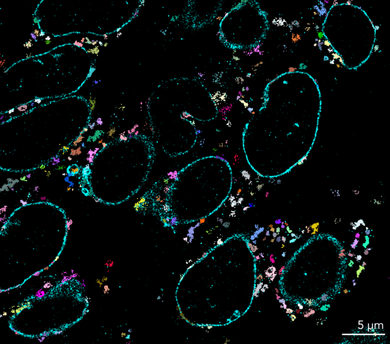
Colorectal cancer tissue clusters
Colorectal cancer tissue sections were stained for lamin (cyan) and TOMM20 (magenta) and imaged with dSTORM. Super-resolution microscopy and the downstream application of clustering algorithms provides an optimal solution to detect nanoscale changes in mitochondrial morphology.

Tubulin dSTORM
dSTORM image obtained with the Nanoimager, showing β-tubulin in COS-7 cells labeled with a primary antibody and subsequently stained with a secondary antibody conjugated to AF647.
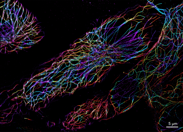
Tubulin 3D dSTORM
Tubulin was labeled with AF-647 in COS-7 cells and imaged using 3D dSTORM on the Nanoimager. Images acquired with Dr. Moreno and Dr. Vivas at University of Washington, US.

Clathrin DNA-PAINT
Clathrin coated pits labeled with a rabbit anti-clathrin heavy chain primary antibody, and detected with the Massive Photonics “massive sdAb-2-plex” kit for DNA-PAINT imaging in fixed COS-7 cells, imaged on the Nanoimager. Localizations are colored by frame index, showing how localizations are overall evenly distributed over the acquisition.
FAQs
Do I need specific laser training or facilities to place the Nanoimager in my lab?
The Nanoimager is a class 1 laser product that can be used in a standard lab, without specific laser training or the need for a dark room. Simply plug it in and image anywhere!
Can ONI’s Nanoimager only do dSTORM imaging?
No, you can do a lot more. The Nanoimager allows you to investigate your sample using different imaging technologies on both fixed or live samples.
Can you use ONI consumable kits with other microscopes?
Yes. Our consumable kits largely simplify sample preparation and these can be compatible with other imaging systems with equivalent specifications. However, all our kits are optimized for use with the Nanoimager.