<p><strong>BrainSTORM Week</strong>: Register now for our upcoming dSTORM webinar series! <strong><span style="text-decoration: underline;">learn more</span></strong></p>

BrainSTORM Week: Register now for our upcoming dSTORM webinar series! learn more

DNA-PAINT

What is DNA-PAINT Microscopy & why is it useful

DNA-PAINT About How it works Usefulness Compatibility
About

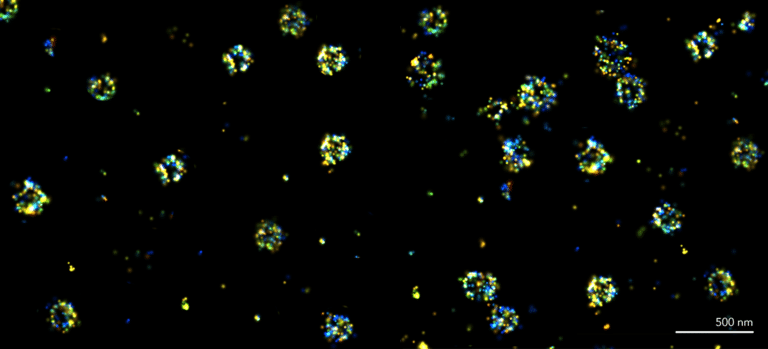
PAINT (Point Accumulation for Imaging in Nanoscale Topography) is a super-resolution microscopy technique that enables the visualization of protein structures smaller than 200 nm by utilizing transient binding events involving DNA strands. By using short, single-stranded DNA (ssDNA) oligonucleotides, known as DNA-PAINT, a docking strand attaches to target molecules like antibodies, while a complementary imager strand linked to a fluorophore captures rapid binding and unbinding events, creating “blinks.”

When illuminated by a laser, these events are recorded over time, allowing single-molecule localization microscopy (SMLM) algorithms to reconstruct super-resolution images with a spatial resolution of about 10-30 nm. This makes PAINT particularly effective for studying subcellular structures and dynamics beyond the diffraction limit of light.

How it works

DNA-PAINT operates on the principle of transient binding between complementary DNA strands to achieve super-resolution imaging. A target molecule, such as a protein, is first labeled with a docking strand of single-stranded DNA (ssDNA), which is immobilized onto the target via a specific antibody or aptamer. Meanwhile, an imager strand, also composed of ssDNA and linked to a fluorescent dye, is kept in solution.

When excited by a laser, the imager strand binds to the docking strand, resulting in a fluorescent “blink” that can be captured by a high-resolution camera. These rapid binding and unbinding events are recorded over time, allowing advanced algorithms to reconstruct precise images at resolutions of 10-30 nm. This process effectively circumvents the limitations of optical diffraction, enabling detailed visualization of subcellular structures.

Usefulness

Significant advantages in imaging compared to traditional fluorescence techniques are offered through DNA-PAINT. Its unique labeling strategy allows for precise targeting and high localization accuracy, while the continuous replenishment of fluorophores prevents photobleaching, a common issue in other methods. The technique also supports multiplexing, enabling simultaneous imaging of multiple targets by using distinct docking and imager strands or sequential flow of new imager strands to new targets over time.

Notably, DNA-PAINT has been demonstrated to visualize up to 124 colors in a single sample through innovative kinetic fingerprinting techniques. Additionally, the quantitative capabilities of DNA-PAINT (i.e. qPAINT) facilitates molecular counting and quantitation, a key aspect to what makes super-resolution microscopy, and more notably DNA-PAINT, a valuable technique.

Compatibility

DNA-PAINT is compatible with various super-resolution microscopy platforms, including the Nanoimager, which is particularly suited for capturing the rapid dynamics of binding events. This versatility allows researchers to integrate DNA-PAINT with other single-molecule localization microscopy (SMLM) techniques, such as dSTORM or PALM, enabling multimodal imaging strategies. Furthermore, the Nanoimager’s microfluidics capabilities streamline the process of exchanging imaging strands, facilitating automated and efficient experiments. Beyond DNA, the platform can accommodate transient interactions involving proteins and glycan-lectin systems, broadening the scope of applications in diverse research fields.

How ONI can help in your DNA-PAINT journey

Sample Preparation
Acquisition
Analysis
Sample Preparation
Acquisition
Analysis

MASSIVE-sdAB ONI 2-PLEX Kit

This kit contains two single-domain antibodies (sdABs) for DNA-PAINT as well as the necessary imager strands and buffers that are required for sample preparation and imaging. Each sdAB can be coupled to a single DNA-PAINT docking site and two sdABs can bind one primary antibody, resulting in two docking sites per primary antibody.

The reagents are tested and optimized for the Nanoimager. Simply add your primary antibodies of interest and you are ready for 2 color DNA-PAINT imaging.

The Nanoimager

Experience the capabilities of our compact benchtop microscope, specifically designed for PAINT imaging and other super-resolution modalities. In addition to PAINT, it supports dSTORM, PALM, Single-Particle Tracking, smFRET, TIRF, and HILO, all within one versatile system. Enjoy seamless integration and exceptional imaging performance tailored for your PAINT applications, enabling you to achieve unmatched resolution and precision in your research!

CODI

Capture fast and transient binding events in PAINT over time with our advanced CODI platform, designed for high-precision localization of single molecules across a large field of view using either TIRF or HILO illumination. Our cloud-based system employs clustering-based algorithms to characterize molecular structures and analyze morphological features, enabling detailed colocalization studies. Enhance your research with unparalleled imaging capabilities that reveal intricate details of dynamic molecular interactions!

Important considerations

How does DNA-PAINT image structures below 20 nm?
How to design your first DNA-PAINT experiment
What are the benefits of multi-modal SMLM in microbiology?
Publications
Technical Content
Videos
BiorXiv, July 2024
High-speed 3D DNA PAINT and unsupervised clustering for unlocking 3D DNA origami cryptography,
Gde Bimananda Mahardika Wisna, Daria Sukhareva, Jonathan Zhao et al.
BiorXiv, March 2024
Multiplexed DNA-PAINT Imaging of the Heterogeneity of Late Endosome/Lysosome Protein Composition,
Charles Bond, Siewert Hugelier, Jiazheng Xing et al.
ACS Sens., January 2023
Precision and Accuracy of Receptor Quantification on Synthetic and Biological Surfaces Using DNA-PAINT,
Roger Riera,  Emmanouil Archontakis,  Glenn Cremers et al.
ACS Nano, June 2023
Mapping Antibody Domain Exposure on Nanoparticle Surfaces Using DNA-PAINT,
Marrit M. E. Tholen Marrit M. E. Tholen Department of Biomedical Engineering, Institute for Complex Molecular Systems (ICMS), Eindhoven University of Technology et al.
Nature Chemical Biology, November 2021
Single-molecule imaging of glycan–lectin interactions on cells with Glyco-PAINT,
Roger Riera, Tim P. Hogervorst, Ward Doelman et al.
ACS Chemical Biology, August 2020
Structure-Based Rational Design of Two Enhanced Bacterial Lipocalin Blc Tags for Protein-PAINT Super-resolution Microscopy,
Liya Muslinkina, Alexey S. Gavrikov, Nina G. Bozhanova et al.

Gallery

Clathrin DNA-PAINT
DNA-PAINT of Nup96-EGFP
PALM-PAINT imaging of RNAP
Nup96 & Nup98 Dual-Color Imaging
Simultaneous Imaging of TOMM20 & Tubulin
Cy3b Tubulin Localization Precision
Clathrin DNA-PAINT
DNA-PAINT of Nup96-EGFP
PALM-PAINT imaging of RNAP
Nup96 & Nup98 Dual-Color Imaging
Simultaneous Imaging of TOMM20 & Tubulin
Cy3b Tubulin Localization Precision