Enter now for your chance to win a 3 month Nanoimager subscription to study your EVs Enter here
Single-molecule FRET microscopy
A Simple Overview
What is single-molecule FRET microscopy?
Single-molecule FRET microscopy (smFRET) is a biophysical technique that enables distances between single molecules to be measured at a scale of 1-10 nanometers. FRET is short for “Förster Resonance Energy Transfer”, a mechanism for describing the transfer of energy between two chromophores.
With smFRET, we utilize the natural phenomenon that occurs between 2 fluorophores, termed donor and acceptor, when they come within 1-10 nm. The emission energy of the donor is transferred to the acceptor which subsequently fluoresces when in close proximity. For FRET experiments donor and acceptors need to be selected carefully so that the donor emission spectrum partially overlaps with the absorption spectrum of the acceptor.
Why is single-molecule FRET microscopy useful?
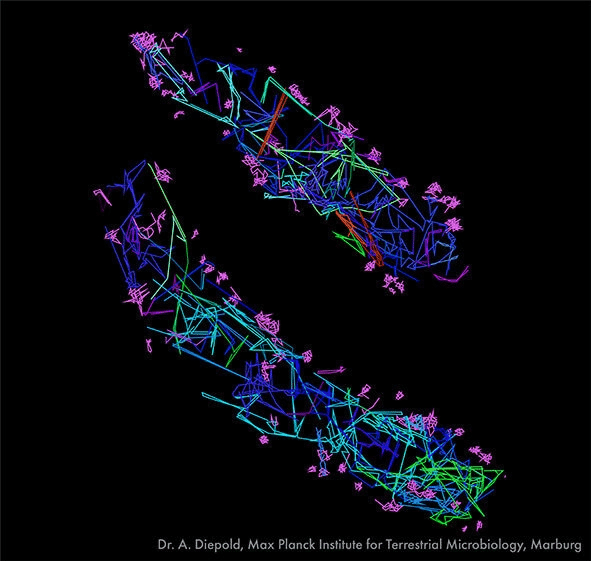
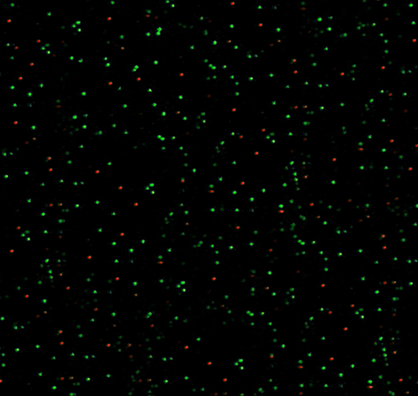
FRET fluorescence can be used in a number ways. Intra- and inter-molecular distances and stoichiometries can measured. In addition, the frequency and duration of binding events can be understood. Going further, smFRET data enables transitional conformational changes to be deduced through qualitative interpretation.
In short, smFRET can be used to quantify and characterise:
- binding events
- intramolecular transitions and dynamics
- specific interactions and complex assembly
- kinetics and dwell times for single molecules
The technique has been usefully employed in many fields including protein folding, DNA conformational change, vesicle fusion and ion channels.

What are the limitations of single-molecule FRET?
The exact distances involved in energy transfer between two fluorophores in a FRET fluorescence event can vary depending on the cell environment and how individual dyes orientate themselves. Fluorophores can bind to different parts of the host protein in a non-standard way and this will affect fluorescence.
However, population averaging can be used to increase accuracy. Furthermore, often to glean significant results, the distance does not need to be ascertained with absolute precision. Movements of individual molecules and dwell times can still be usefully obtained.
Which microscopes are capable of single-molecule FRET?
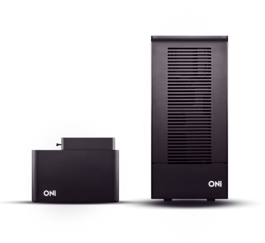
The Nanoimager is the only commercially available microscope capable of carrying out single-molecule FRET microscopy. The Stage Top Incubator complements this by providing stable environmental conditions for live-cell studies, further enhancing the capabilities of single-molecule FRET. It comes with our NimOS software, which interprets the smFRET imaging data and enables reactions to be followed in real-time. With the large field of view, thousands of FRET fluorescence events can be traced in a single acquisition, speeding up the process and improving the accuracy of results. Individual FRET traces can be plotted and population averages as a whole can calculated.
Read about other super-resolution microscope techniques and illumination modes available on the Nanoimager microscope. Alternatively, read the application for revealing molecular mechanisms and interactions by single-molecule FRET.