Upcoming Webinar – Small dyes, big impact: Enabling gentle super-resolution imaging in live cells register now>
Discovery Kit™: dSTORM in cells 2
The ultimate kit to prepare your samples for super-resolution

From basics to brilliance
Prepare your samples for dSTORM super-resolution imaging with ease
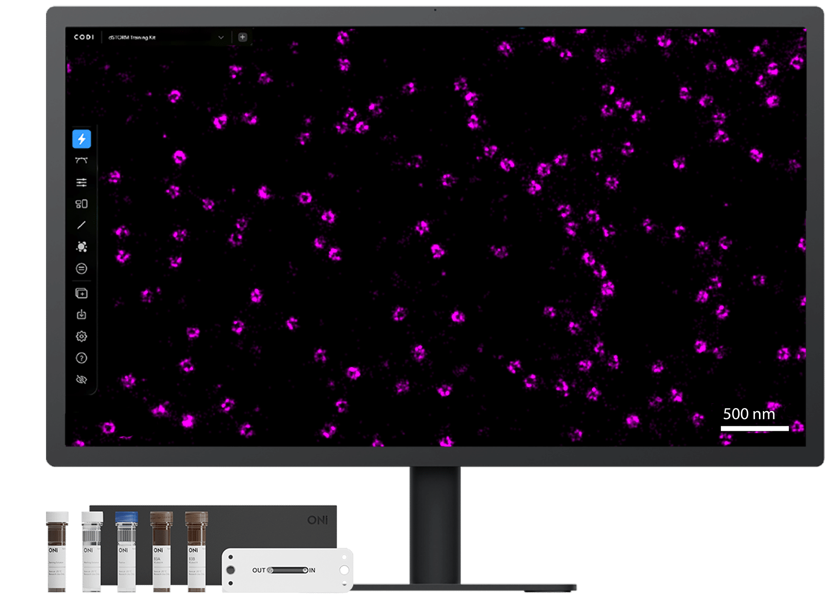
The ONI Discovery Kit™ for dSTORM in cells 2 imaging provides a modular workflow for immunofluorescent labeling in cultured cells, which allows you to confidently detect extra and intracellular proteins in two channels with 20 nm resolution and high sensitivity in your own samples.* You provide the cells and custom antibodies, we provide the rest!
The kit enables you to:
- Label targets of interest with your own primary antibodies
- Use highly specific anti-mouse, rabbit and rat secondary antibodies conjugated with best-in-class dSTORM fluorophores
- Optimize key sample preparation steps, such as fixation, to preserve biological structures and avoid artifacts
- Perform 2-color super-resolution dSTORM imaging in various cell types
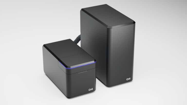
- Obtain images with 20 nm resolution using ONI’s Aplo Scope & Nanoimager
- Use ONI’s CODI platform for cluster-based data analysis to extract novel nanoscale cellular information
*Reagents have a shelf life of minimum 2 two months and up to 6 months.

Which kit is right for me?
Prepare your samples for dSTORM super-resolution imaging with ease
Discovery Kit: dSTORM in cells 2
The standard kit for intracellular proteins

The standard Discovery Kit: dSTORM in cells 2 helps you prepare cells for dSTORM imaging with ONI’s optimized reagents and protocols, allowing you to save time and obtain robust super-resolution data in a fast and simplified manner. We recommend this kit for researchers studying intracellular non-membrane proteins. Exceptions are structural proteins, such as tubulin and actin, which are optimally fixed with the stronger fixative.
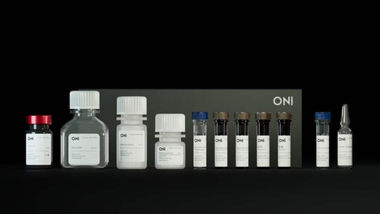
What’s in the kit?
PFA-based fixative: Ready-to-use PFA is provided in a tear-off amber glass vial to avoid polymerization or deterioration. It is also used for a post-fixation step to cross-link antibodies to targets.
Blocking buffers: Simultaneous blocking and permeabilization to save time and permeabilize cells all the way to the nucleus.
Fluorescently-labeled F(ab’)2 secondary antibodies:
Smaller than full length IgG to reduce linkage error. Choose two antibodies from three different options (rat, mouse or rabbit) with either Cy3B or AZDye™647, the best fluorophores for dSTORM for 2-channel imaging.
dSTORM imaging buffer: Our dSTORM imaging buffer provides excellent blinking and anti-bleaching activity.
Comprehensive protocol: Guides you through sample preparation, includes troubleshooting notes and advice to optimize antibodies against your protein of interest.
Discovery Kit: dSTORM in cells with Strong Fixative 2
The kit to try for membrane & structural proteins

PFA can be insufficient to cross-link many membrane proteins, which remain mobile¹. This can lead to antibody-induced protein clustering²,³. The Discovery Kit: dSTORM in cells with Strong Fixative 2 immobilizes membrane proteins using a combination of PFA and glutaraldehyde. This better preserves the native structure and enables you to confidently measure membrane protein clustering. Because glutaraldehyde can disrupt some epitopes this kit provides both fixatives for you to compare and choose the best fixative for your protein of interest.
What’s in the kit?
All components listed in the standard Discovery Kit: dSTORM in cells 2
PFA-based fixative and Fixation Supplement for strong fixation: Ready-to-use PFA is provided in a tear-off amber glass vial to avoid polymerization or deterioration. Extra PFA is provided for a post-fixation step so antibody labeled samples can be stably stored in the fridge. The Fixation Supplement (glutaraldehyde-based) helps minimize residual mobility and avoid artifacts.
Quenching buffer: A quenching reagent is supplied to fully reduce the glutaraldehyde to minimize autofluorescence and non-specific antibody binding.
REFERENCES
Tanaka K, Suzuki K, Shirai Y et al. Membrane molecules mobile even after chemical fixation. Nat Methods. 2010; 7, 865–866. https://doi.org/10.1038/nmeth.f.314
Stanly TA, Fritzsche M, Banerji S, García E, Bernardino de la Serna J, Jackson DG, Eggeling C. Critical importance of appropriate fixation conditions for faithful imaging of receptor microclusters. Biol Open. 2016; 5(9): 1343-50. doi: 10.1242/bio.019943.
Werner C, Sauer M and Geis C. Super-resolving Microscopy in Neuroscience. Chem. Rev. 2021; 121 (19): 11971–12015.
Features & Benefits
50 samples with 1 kit
Volume provided is for 100 µL per sample. Protocol validated on uncoated and poly-L-lysine coated surfaces.
Optimize fixation & labeling for your targets
Use either the standard Discovery Kit: dSTORM in cells 2 or the Discovery Kit: dSTORM in cells with Strong Fixative 2 to fix and label your intra or extracellular targets of interest across a range of cell lines.
Choose secondary probes for 2-color imaging
Three different species of secondary antibody are offered as part of the dSTORM Discovery Kit, from which you can choose two conjugated to either Cy3B or AZDye™647.
dSTORM imaging using the Nanoimager or Aplo Scope
Stained cellular targets, including those expressed at low levels, can be detected using the Nanoimager or Aplo Scope, ONI’s desktop super-resolution microscope, which allows simultaneous or sequential imaging of targets stained with Cy3B or AZDye™647 secondary antibodies.
Analyze dSTORM with ONI’s CODI platform
Extracting nanoscale morphological information from the stained structures is possible with our cloud-based analysis platform, CODI, which includes clustering and counting to assess protein distribution in cells.
The proof is in the data
ONI’s in-house team of experts has tested a range of protein targets with the dSTORM Discovery Kit, including:
Intracellular organelles: mitochondria early and late endosomes, lysosomes
Structural proteins: tubulin
Nuclear proteins: histones, lamina
Surface receptors including those at low expression: CD3, CD28, PD1
Tested in a range of cell types including human cancer cell lines, primary neurons, T cells, and mouse cell lines
* These images were acquired with the Discovery Kit™: dSTORM in cells 1

Lysosomal LAMP-1 (magenta) and tubulin (yellow) labeled with anti-rabbit CF583R and anti-rat AZ647 in U2OS cells.

Mitochondrial TIMM44 (magenta) and TOMM20 (green) labeled with anti-mouse CF583R and anti-rabbit AZ647 in U2OS cells. Zoomed images highlight the power of dSTORM to distinct the two membranes at nanoscale level.
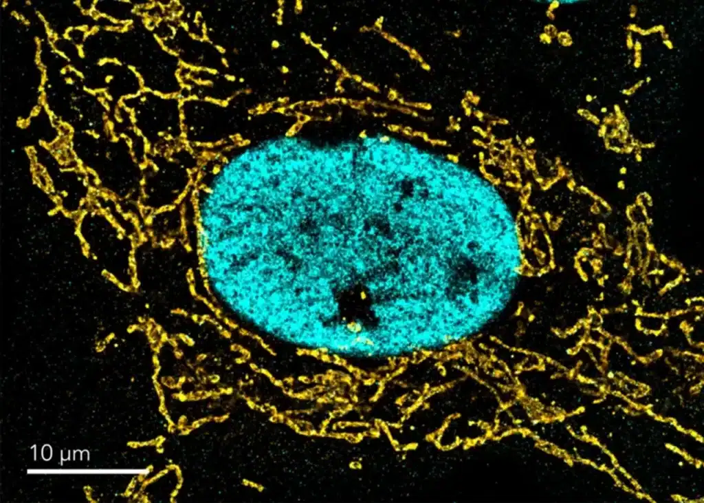
Histone H3K27ac (cyan) and mitochondria TOMM20 (gold) labeled with anti-rabbit CF583R and anti-mouse AF647 in U2OS cells.

Neuronal synaptic markers Bassoon (magenta) and Homer1 (cyan) labeled with anti-mouse AZ647 and anti-rabbit CF583R (cyan) in primary rat cortical neurons.
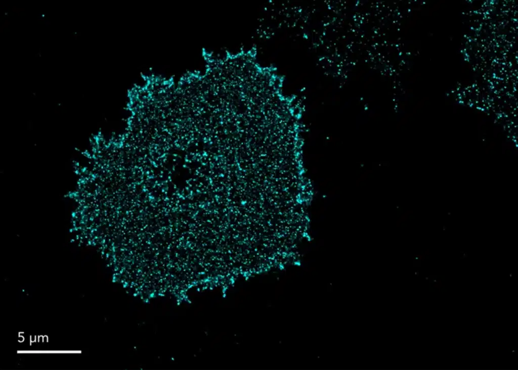
PD-1 labeled with anti-mouse AZ647 (cyan) in primary T cells.

Lysosomal LAMP-1 (magenta) and endosomal CD63 (green) labeled with anti-rabbit CF583R and anti-mouse AZ647 in U2OS cells.
Ready to purchase?
Join an upcoming Live from the Lab
Join an upcoming Cell Imaging Training
Join our Cell Imaging Training with the ONI Discovery Kits™, where you’ll learn to use a modular workflow for immunofluorescent labeling in cultured cells. This training will guide you through detecting extra- and intracellular proteins in two channels with 20 nm resolution and high sensitivity. You’ll provide the cells and custom antibodies, while we supply the reagents and tools to achieve precise and reliable results. Gain hands-on experience with the kit, from sample preparation to image analysis, and optimize your workflow for high-quality cellular imaging.