Run CODI Locally With CODI Offline learn more
ONI Achieves First-Ever Molecular Resolution in a Fully Automated Optical Imaging Workflow
Author: Carolyn Hartmann
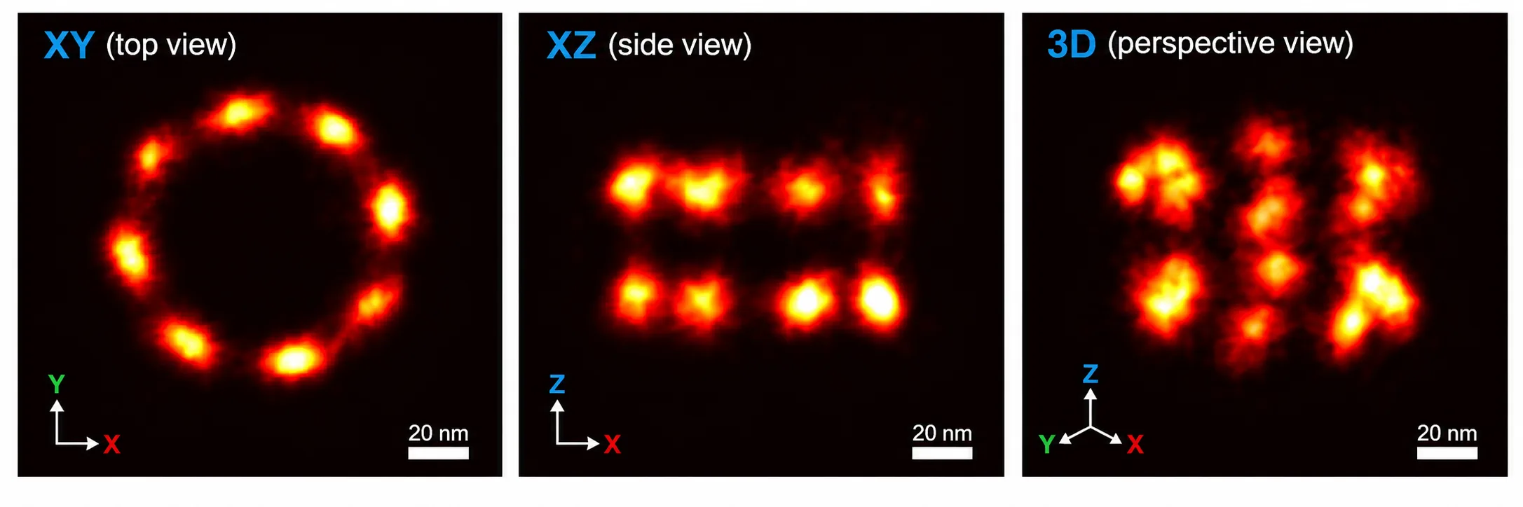
Image: Automated 3D RESI imaging of Nup96 using Aplo Flow and Aplo Scope, shown in XY, XZ, and 3D perspective views.
FOR IMMEDIATE RELEASE
New research from ONI demonstrates how an automated RESI workflow can distinguish protein structures just nanometers apart while achieving sub-nanometer localization precision.
SAN DIEGO, CA and OXFORD, UK, September 16, 2026
ONI, the leader in super-resolution microscopy and single-particle analysis, has unveiled new research that gives scientists an extraordinarily detailed view of how proteins are organized at the molecular scale. Researchers at ONI used a technique known as Resolution Enhancement by Sequential Imaging (RESI), first described by Reinhardt et al. in Nature in 2023, within a fully automated workflow demonstrating sub-nanometer localization precision.
The implications are significant for drug discovery. By combining molecular-scale detail with automation and higher-throughput imaging, researchers can study more samples, compare more biological conditions, and gain a clearer view of how therapies actually work inside cells. This information can help scientists better understand mechanisms of action, identify promising drug targets, make more informed decisions earlier in development, and ultimately help shorten the path from discovery to the clinic.
Understanding complex biology often requires more than knowing whether a protein is present or absent. Researchers also need to understand precisely where proteins are located within a cell, how closely they sit together, and how they are organized relative to neighboring molecules. These spatial relationships can provide important insight into protein interactions, receptor organization, antibody binding, disease mechanisms, and therapeutic function.
For years, getting this level of molecular detail has come with a major tradeoff: complexity. Techniques like RESI require repeated rounds of imaging, careful alignment, tightly controlled conditions, and significant hands-on expertise. Even small changes in temperature, vibration, focus, or sample position can affect the result. That has made these experiments difficult to run consistently, hard to scale, and challenging to bring into higher-throughput research environments.
ONI’s new research demonstrates that the complete RESI imaging workflow can be automated while maintaining sub-nanometer localization performance. A single field of view can take five to seven hours to acquire, making automation essential for reducing hands-on work, improving consistency across long experiments, and enabling researchers to scale these workflows to larger, higher-throughput studies. That combination of molecular-scale resolution and automation also creates the potential to generate larger, more consistent imaging datasets from experiments that have historically been difficult to scale, opening new opportunities for data-intensive discovery and AI-driven biological research.
Unlike many emerging imaging advances, this capability is not confined to the research lab. The underlying technology is commercially available today exclusively through ONI and is already being used in biopharma.
Using Aplo Flow an automated fluidics platform integrated with the Aplo Scope super-resolution microscope, ONI automated the repeated fluid-exchange and imaging steps required across multiple RESI cycles. The result: sub-nanometer localization precision and the ability to distinguish neighboring protein structures just 10 nanometers apart.
The workflow achieved 0.97 nm lateral localization precision, allowing researchers to map molecular positions with enough accuracy to reconstruct nanoscale protein organization in three dimensions.
“RESI can reveal molecular organization at an extraordinary level of detail, but repeating the fluid exchange and imaging steps across multiple cycles makes these experiments technically demanding,” said Ricardo Nunes Bastos, PhD, Chief Scientific Officer, ONI. “By automating those cycles as a coordinated workflow, we were able to preserve the precision needed to distinguish protein structures only 10 nanometers apart while reducing the hands-on complexity of the experiment.”
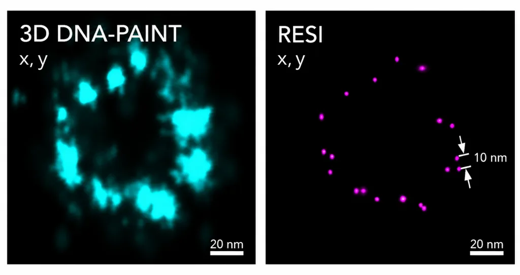
Image: Automated 3D RESI imaging of Nup96 resolves protein structures 10 nm apart.
See the data in action in ONI’s on-demand webinar.
About ONI
ONI is a private life sciences tools company that transforms therapeutic discovery with accessible, high-performance super-resolution microscopy. Its backers include ARCH Venture Partners, Casdin Capital and Oxford Science Enterprises (OSE). The company’s first product, the Nanoimager, introduced the world’s first desktop platform for single-molecule imaging, offering 15 nm resolution to study individual molecules in living cells, tissue, and nanoparticles. ONI’s latest innovation, the Aplo Scope, is a compact, end-to-end imaging platform that delivers unmatched clarity, precision, and speed. Designed to empower researchers, the Aplo Scope provides real-time insights into complex biological systems, enabling breakthroughs in drug delivery, biomarker discovery, and antibody therapeutics. For more information, visit oni.bio. Follow ONI @oniHQ and LinkedIn.
For media inquiries, please contact:
Carolyn Hartmann
Director of Product Marketing, ONI
chartmann@oni.bio