Introducing: ONI’s Next-Generation End-to-End Solution for EV Research Learn More
Tracking Single Molecules and Vesicles in cells
Challenge
Understanding particle dynamics
Understanding the dynamics, trajectories and interactions of single molecules and vesicles in cells presents a fundamental challenge in cell biology. With single-particle tracking it is possible to study the dynamics of the plasma membrane, of transcription, of protein-protein interactions and also the influence of external factors such as drugs on these mechanisms. However, an instrument that is fast enough to record these processes and has analysis that is simple to navigate is critical.

Solution with the Nanoimager
Single-particle tracking
The Nanoimager offers a complete solution for tracking single particles and vesicles. The option to heat the microscope to 37℃ allows live-cell imaging and the simultaneous two color acquisition offers the incredible possibility to localize and track two different types of molecule or vesicle at the same time. The NimOS software provides dedicated single-particle tracking analysis, automatically detecting either molecules or vesicles and tracking them over time. The diffusion coefficients and measurements of the size and number of particles (for tracked particles in solution) is summarized instantly, for single or groups of tracks, with the option to export results in a convenient CSV format.
By following the trajectories of fluorescently labelled EVs in solution, the Nanoimager can apply nanoparticle tracking analysis (NTA) style sizing and quantification of vesicle populations. It also uniquely offers the ability to size EV populations in addition to visualizing and tracing their dynamics in a cellular context. The case study below illustrates an excellent example of this multi-method approach to understanding the role of EVs in cancer.
![]()
Simultaneous dual-color tracking of extracellular vesicles, red color is Cd81-RFP, yellow is EV-marker-GFP (scale bar: 2µm)
Case Study
Single-particle tracking with TRP channel proteins
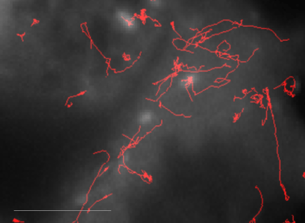
With single-particle tracking, we can understand the dynamics, trajectories and interaction of single molecules and vesicles in cells. TRP channel proteins were tagged with mCherry and expressed in HEK cells. They are transported to the plasma membrane in vesicles, which are visible as the highly dynamic bright fluorescent spots moving throughout the cells. In the image, the trajectories followed by the vesicles were mapped out using the NimOS software tracking feature.
Read more about the Nanoimager’s applications for viral particle imaging and studying exosomes and microvesicles.

